News

Explaining Brain Injury to Your Child

Chapter Four, The Caregiver’s Journey on tbi.cemmlibrary.org


Having a parent with a TBI can be frightening for a child who looks to them to provide strength and safety.
A parent with TBI may no longer act the same as they did before the injury. They may be angry, depressed, or uncertain. They may not be able to speak or walk, and they may not be able to do all the same things they did before they were injured (for example, go to work, pick up the kids from school, play on the playground). You can help your children by explaining TBI in a way that they can understand, or you can ask a healthcare provider to talk with your children.

As a result of the changes caused by the injury, the special parent-child bond that existed previously has probably changed, at least to some degree.

Children may be confused and upset about what is going on. This could be due to worry about a parent’s condition or concerns about changes in their parents’ relationship. It could also be due to financial strains, or simply adjusting to the new “normal” (in other words, the new way of life following the TBI). Be sure to communicate with your child that they are not to blame for the TBI – many children will assume they somehow contributed to the injury and will carry some sense of responsibility for it. If your child appears to be anxious or depressed for a long time, or they begin taking on risky behaviors, seek professional help.

It is important to recognize that your children are grieving, just as you are – they are grieving the loss of the parent’s former “self” and the abrupt changes that have taken place. They may withdraw from social activities with peers, have mood swings, become withdrawn or disruptive, do poorly in school, and show other behavioral problems.

Children also need time and space to be kids. Allow them time to think, play, talk, or just hang out – don’t smother them with too much information or attention over the injury – but be sensitive to their questions and concerns. Build new family routines, and keep an eye out for signs that your child is not coping well.

Some children may need to take on some caregiving tasks for the parent or for younger children in the family. Children who care for parents or other relatives may experience conflict over the reversal of roles between parent and child. Others will find it helpful for them to play a supportive role.

Make sure any tasks that your child takes on to help around the house or with caregiving activities – household chores or meal preparation, for example – are suitable for their age. Strive as much as possible to find other adults to help you, rather than relying on your children to play a major caregiving role.


How Can I Tell My Child about TBI?

It is difficult to explain TBI to a child. Yet it is vital to tell your child what is going on. Some adults try to protect children from the truth because they think they are too young to understand. Children of almost any age are aware that something is wrong and they want to know what is happening.

Communicate in an age-appropriate way what has happened to the service member or veteran with TBI. Protecting your children by withholding information may backfire. Children have active imaginations that may create a scenario worse than reality.

How you tell your child about TBI depends on the age of the child. Here are some suggestions for how to explain TBI to a child:

  • Use Language a Child can Understand: The brain is similar to the command station of a spaceship. If a meteorite hit the command station, the crew would not be able to control what the spaceship does. If the brain is hurt, it may send out the wrong signals to the body or no signals at all. A person with TBI may have a hard time walking, talking, hearing, or seeing.
  • The brain is the body’s computer. When it gets injured, it doesn’t “boot up” properly, runs slower, has less memory, and may not perform all of its normal functions.
  • A cut may take a few days to heal, and a broken bone will usually heal and be as good as new in a few weeks or months. Getting better after a brain injury can take several months or even years, and sometimes the person may not get 100 percent better.
  • Even though the person with the injury may look the same, they may still be injured. These injuries might include having a hard time paying attention or remembering what you told them. They may get tired easily and need to sleep more than usual. They may say or do things that seem strange or embarrassing. They may get angry and shout for no apparent reason.
  • Explaining Anger Changes: Many people with a TBI develop anger as a direct effect of the damage to the brain. In other words, the parts of the brain that normally stop angry flare-ups and feelings have been damaged and don’t do their job as well. The parent with TBI may be mad because they can’t do the things they used to do. Their feelings may be hurt because others treat them differently than before the injury.
  •  
    Brain injury changes people. These changes can be confusing. Try to remember that the changes you see are caused by the brain injury. You can still love and care about the person regardless of the brain injury.
     

    CLICK HERE to read the original article


    The Benefits of Smiling

    By Jennifer, spindpals.com, July 16, 2020

     
    How many times have you smiled today?  If you’re an average adult, you’ll smile 20 times today.  If you’re a really happy adult, you’ll smile 40-50 times today.  That sounds decent, until you compare it to the number of smiles a child expresses each day: 400!
     
    Why do we smile and how does it affect our brain?  Here’s the sequence of events:

  • You feel happy
  • Your brain sends a signal to cranial nerve VII, which then triggers the face’s zygomaticus major muscle (the one responsible for lifting the corners of your mouth) and the obicularis occuli muscle (the one around your eyes)
  • You smile
  • A positive feedback loop is initiated and a signal is sent back to your brain, releasing dopamine and serotonin, and reinforcing positive emotions
  •  
    Other benefits of smiling:

  • It lowers the stress hormones of cortisol and adrenaline
  • It boosts the immune system by increasing gamma interferon (a protein that fights viruses), B-cells and T-cells (the white blood cells needed to create antibodies)
  • What about fake smiles?  It turns out that fake/”social” smiling enough can actually bias your brain to thinking you are genuinely happy due to the positive feedback loop.

    —>FUN FACT:  A single smile can stimulate neurotransmitters as much as 2000 bars of chocolate, but without the sugar crash and stomach ache!
     
    When you next pass someone on the street, flash your pearly whites.  You’ll make them feel good and cheer yourself up as well.

    “Sometimes your joy is the source of your smile, but sometimes your smile can be the source of your joy” ~Thich Nhat Hanh

    CLICK HERE to read the original article


    My Tech Tools for TBI


    By Alexander Rostron, August 25, 2020

     
    After my traumatic brain injury (TBI), I suffered from a large drop in mental capacity.  I could not remain focused for even moderate lengths of time and forgot the majority of the content that I had just consumed.  

    Not only that, whenever I attempted focused work, I was so mentally and physically tired after doing something as simple as reading that I needed to take a nap.  The limited runs of mental stamina prompted me to explore different tools and systems for assistance.

    Below are three apps I find useful:

    Notability App (iOS, Mac)

    I use Notability to write notes (handwritten and typed), take photos and record audio from class and during medical appointments.  My favorite feature is the ability to capture audio while handwriting notes and highlighting key terms with the Apple Pencil.  

    While reviewing my class notes to study for a test, I realized that the audio recorded more information than I was able to put down during the lecture.  For example, a professor gives an example of the definition they had just explained.  I was only able to write out the definition, before the professor started on a different part of the lesson.  The audio recording left me more information than my writing speed could handle.

    Google Calendar

    I use Google Calendar to track the scheduled appointments I have.  My favorite part of that utility is the option of adding details to each event, such as the address, a notification to remind me an hour before, and a space for a custom description.  Here is where I list important details about what needs to be brought and prepared for the appointment.  I have used it to track things I need to bring and do before each appointment.

    Forest Pomodoro Timer (iOS, Mac)

    I use Forest to practice the Pomodoro technique for working on tasks that take intense focus, while giving scheduled breaks for a mental recovery.  It is a timer that stops if you use your phone.  This is to practice only studying for a set amount of time, with an alert after as a reminder to physically get up from where I’m studying and take a very short break.  Studies have shown that after 25 minutes of intense mental focus, human ability to retain information gets severely diminished.  This is a tool I consistently use when I am completing a task with a lot of paperwork and takes a lot of time and mental energy, such as writing essays or personal record-keeping.

    These 3 tools act as such a critical part of my life post-TBI.  The Notability app catches all of the information that I cannot record at the time of, and helps support my memories with the tools it gives to type, write, and emphasize certain information.  The Google Calendar system helps give me a clear layout of the events in the week ahead of me and contains a place for each small and important detail.  The Forest app helps me pace myself through large blocks of work with small checkpoint breaks to ensure that I don’t drain my mental battery and a chance to reflect about what info I had just absorbed.

    These are the systems that assist me with the mental stamina and memory recall struggles specific to my traumatic brain injury. If you have tools that you would like to add to this list, I would love to hear about what you have used to support your life post-TBI. If you would like to hear more about one of these tools please let me know.  If you have any comments, questions, or feedback please feel free to reach out to me at arroston@gmail.com.

    CLICK HERE to read the original article


    Alcohol and Brain Injury

    After a TBI or Stroke the brain can become more sensitive to the effects of alcohol. This can cause cognitive problems that impact memory, mobility, and speech. It can also cause someone to feel fatigued and unwell. What can be expected after your recovery?


    By Fred at Spindpals.com, July 18, 2020


    Whether we’re on holiday abroad or enjoying the Christmas festivities, an alcoholic drink tends to not be too far from reach for many of us.

    But after a brain injury, the body’s tolerance to alcohol is greatly reduced, and many survivors find that they are no longer able to enjoy alcohol in the same way as they did before their injury. The reduced tolerance to alcohol means that many effects of brain injury are exacerbated after drinking, such as memory problems, mobility issues, speech and fatigue.

    Remember you should always discuss with your medical practitioner your particular condition to understand what the impact would be on yourself. Never take alcohol without their approval and guidance.

    AUTHOR: “It is clear that there is an uneasy relationship between alcohol and brain injury. Survivors are often faced with the challenge of balancing a desire to enjoy the social life they had before they sustained their injury with the acceptance that alcohol now affects them in a different way.”

    We asked brain injury survivors to tell us about how their relationship with alcohol has changed.

    For some, the enjoyment of drinking is simply outweighed by the effects caused.

    “I don’t drink anymore,” said Louise Fry. “I couldn’t drink to start with because of meds, but now? It just hits me too hard.”

    Janet Creamer agreed: “Drinking is now a no-no. Just one alcoholic drink does awful things to my brain. It feels like I’ve drunk way too much and I get that spaced out feeling.”

    Others, like Giles Philip Hudson, have found that being advised by doctors to no longer drink has actually been a blessing in disguise.

    AUTHOR: “After sustaining my brain injury and spending over four months in hospital, doctors advised me not to drink alcohol. During this time I found I no longer needed to drink alcohol to make me feel good or enjoy myself. I certainly don’t need the headaches it causes.”
     

    Enjoy a drink at home with family and friends

    Naturally, many people want to continue to be able to enjoy a drink every now and then, particularly at social gatherings. But what if going to the bar is too daunting a prospect?

    Home drinking is increasingly popular For some, staying in allows them to enjoy a drink without some of the challenges of being in a busy, crowded and noisy pub or bar.

    “Since my disability I do not feel comfortable going into a bar as I may find it hard to use the restrooms,” said one member of the community, “so my drinking is done in my home.”

    Patricia Nugent on Facebook agreed: “We tend to drink at home so it is easier and less stressful to moderate intake,” she said.

    If you are choosing to drink at home, it’s important you monitor your intake carefully.
    Here’s some useful advice for home drinkers:

      1. Keep track of how many units you’re consuming
      2. Use smaller glasses
      3. Use proper spirit measures to avoid inadvertently pouring yourself a double or triple measure
      4. Eat as you drink
      5. Invest in a good bottle stop to make that bottle of wine last longer

    Out and about

    For others, however, a good night out is still a must! If that’s the case, then planning ahead can be the key to the success of the evening.

    “I don’t go out much, once every two months,” said Michelle Richardson. “But it’s lovely to have some drinks and let my hair down and forget how challenging recovery is for a while.

    AUTHOR: “I do have to prepare for a night out by having an afternoon snooze.”

    If you do want to enjoy a night out on the town with friends, here are some more top tips:

      ▪ Don’t drink on an empty stomach and check your medication allows you to drink
      ▪ Make sure your friends know about your brain injury, lowered alcohol tolerance levels, and any other issues such as an intolerance to noise
      ▪ Drink water between alcoholic drinks and avoid getting into rounds

    Alcohol-free alternatives

    Of course, not drinking alcohol doesn’t mean you can’t still go out to pubs and bars.

    “My husband has been told he can’t drink alcohol,” said Amanda Hopkins. “So, as he is a real ale drinker, we made a pact to still go to country location but to just check out ‘alcohol-free’ ales and to become connoisseurs of the growing ‘alcohol-free’ ranges that are now appearing from many microbreweries.

    “It won’t be quite the same but we hope it will be a bit of fun tasting them.”

    AUTHOR: Kathy M agreed: “I sometimes have a non-alcoholic beer shandy so I feel like I am having a pint and I’ve discovered things like elderflower cordial with soda. There’s nothing wrong with ordering a fancy coffee or mocktail either.”

    Drinking alcohol after Stroke

      ▪ Drinking too much alcohol contributes to a number of risk factors for stroke, including high blood pressure.
      ▪ Alcohol can interfere with the medicine you take to reduce stroke risk.
      ▪ Your doctor can advise when it is safe for you to start drinking alcohol again and how much alcohol it is safe for you to drink.
      ▪ Healthy men and women should have no more than two standard drinks a day, and no more than four standard drinks on any one occasion.

    Alcohol and stroke risk

    Drinking too much alcohol contributes to a number of risk factors for stroke. If you have already had a stroke or transient ischaemic attack (TIA), you can help reduce your risk by only drinking a safe amount.

    High blood pressure is the biggest risk factor for stroke, and drinking too much raises your blood pressure. Atrial fibrillation, which is a type of irregular heartbeat, can be triggered by too much alcohol.

    Diabetes and being overweight also increase your risk of having a stroke. Both of are linked to alcohol consumption.

    Alcoholic drinks are also high in calories with little nutritional value. Reducing the amount you drink will support you to maintain a healthy weight.

    Hemorrhagic stroke and alcohol

    A hemorrhagic stroke is caused by a break in the wall of a blood vessel in the brain. If you have had a hemorrhagic stroke, you must not drink alcohol for at least three weeks after your stroke. Ask your doctor when it is safe to start drinking alcohol again.

    Drinking alcohol and your medication

    Alcohol could interfere with the medicine you take particularly, blood-thinning medicine such as Warfarin. Discuss with your doctor about whether it is safe to drink alcohol while taking any medicines.

    Consuming alcohol safely if your doctor clears you to

    The Guidelines for Alcohol Consumption gives advice about safe amounts of alcohol.

    AUTHOR: Remember, the Guidelines are for healthy people. Talk to your doctor about whether it is safe for you to drink at all, and whether the amounts in the Guidelines are safe for you.

    The Guidelines state that healthy men and women should have no more than two standard drinks on any day and if you go out, no more than four standard drinks on any one occasion.

      ▪ For spirits with 40 % ABV, a standard drink is 30 mls (1.5 fl oz)
      ▪ A 285 ml (10 fl oz) glass of 3.5% ABV beer is about 1 standard drink.
      ▪ 100 ml (3.5 fl oz) of wine or champagne is approximately one standard drink, however this varies between types. Keep in mind most glasses of wine served in restaurants and bars are more than 100 ml (3.5 fl oz).
      ▪ Always check the label on the bottle to find out how many standard drinks you are having.
      If you find you are tempted to go over your safe limits learn strategies to help you keep to them.

    Strategies to reduce your drinking

    Write down how many drinks you have to see how much and how often you drink.
    If you find that you are drinking more than is safe, try these tips:

      ▪ Drink water when you are thirsty rather than alcohol.
      ▪ Sip your drink slowly. Put down the glass after each mouthful.
      ▪ At social occasions, make every second drink a non-alcoholic beverage. Choose something like a sparkling water rather than a sugary drink.
      ▪ Try low-alcohol alternatives such as light beer.
      ▪ Opt out of ‘shouts’. Drink at your own pace. If you cannot avoid buying a round, get yourself a non-alcoholic drink.
      ▪ Avoid salty snacks such as potato chips or peanuts. These make you thirsty and more inclined to drink quickly.
      ▪ Set goals such as not drinking alone and have at least two days without alcohol each week.
      ▪ Do not drink on an empty stomach. A full stomach slows the absorption of alcohol

    Brain Injury affects different people in different ways. There is no one size fits all. For some, their relationship with alcohol will be over. For others, a moderate consumption can be tolerated. Complete your recovery and then discuss with your medical practitioner your options. Do not make any decisions without consulting them first.

    This post is shared from the Stroke Association AU and Headway UK websites
     
    CLICK HERE to read the original article
     

    Pictured is a high-fidelity map of physician-scientist Nico Dosenbach’s brain while his dominant arm was in a cast for two weeks. The red and yellow areas of the MRI image represent previously undetected brain pulses. Dosenbach and colleagues at Washington University School of Medicine in St. Louis found that disuse of an arm causes the affected brain region to disconnect from the rest of the brain’s motor system within two days. However, spontaneous pulses maintain activity in the disused circuits until the region becomes active again when mobility is regained.


    Previously undetected brain pulses may help circuits survive disuse, injury

    Research may lead to treatment advances for patients with immobilizing illness, injury

    By Washington University in St. Louis, June 15, 2020


    To study brain activity, Nico Dosenbach, MD, PhD, an assistant professor of neurology at Washington University School of Medicine in St. Louis, wore a fiberglass cast for two weeks.

    A neuroscientist’s neon pink arm cast led him and fellow researchers at Washington University School of Medicine in St. Louis to discover previously undetected neuronal pulses in the human brain that activate after an immobilizing illness or injury.

    The pulses appeared on MRI scans used to measure brain activity of the neuroscientist and, later, two additional adults whose arms were in casts. The researchers compared those MRI images with scans of the scientists before and after their arms were put in casts.

    The scans showed that the brain’s main circuits responsible for movement in specific areas of the body disconnected within 48 hours of a person wearing a cast that encumbered movement in such an area. Also during this time, “disuse pulses” emerged to maintain neural activity and allow the main motor circuits to reactivate if and when mobility was restored through physical therapy.

    The findings, published online June 16 in Neuron, offer clues to how the brain’s billions of neurons — cells that transmit nerve impulses — can rewire and restore pathways after injury or illness. Understanding just what is behind this resiliency may lead to new therapies for people with broken limbs or recovering from strokes or other immobilizing conditions.

    “Many scenarios exist in neurology in which a person doesn’t use an arm or a leg and, consequently, related brain circuits for an extended period of time,” said senior author Nico Dosenbach, MD, PhD, an assistant professor of neurology. “In offering the best care to patients, it’s important to understand specifically what changes occur in brain function. Accurate understanding and mapping of these circuits may lead to advancements in treating patients who have lost use of their limbs.”

    In 2015, Dosenbach — also an assistant professor of occupational therapy, of pediatrics, of radiology and of biomedical engineering — wore the pink cast for two weeks despite the fact he had no injury requiring one. He aimed to collect high-quality data using brain-imaging techniques to evaluate neural networks that control movement.

    Many of the patients Dosenbach treats at St. Louis Children’s Hospital suffer from conditions that limit mobility and cause them to favor one side of the body. A common treatment is constraint-induced movement therapy, also known as forced-use therapy, which immobilizes the dominant arm with a cast, forcing the child to use the impaired arm.

    “My goal was a better understanding of what my patients experience during therapy, although I acknowledge it’s more difficult for them because of their disabilities,” Dosenbach said.

    He also wanted to pinpoint a timeline of when individual neural changes occur. Commonly, scientists gather MRI data from dozens of people and average it. “But I did not want to do that because everyone’s brain is anatomically different, and when MRI data are averaged, it all blurs together,” Dosenbach said.

    So Dosenbach decided to wear a fiberglass cast on his dominant, right arm. It stretched from his fingertips to just below his shoulder. It was pink, the favorite color of his daughter, Maike, then age 2.

    He wore it during the hot, humid summer. It itched. It was awkward. He had to learn how to change a diaper with one hand. 

    Every day, he arose predawn to lie stiffly for 30 minutes for a resting-state functional MRI. He did this for the two weeks his arm was in the cast, as well as for the two weeks before and after.

    During the six weeks, Dosenbach also wore accelerometers on both wrists to track the motor strength of his arms while performing basic tasks such as writing and moving objects.

    “It wasn’t terrible, just unpleasant,” Dosenbach recalled. “But immediately, I noticed my right arm got worse, and my left hand got stronger. It was much faster than any of us expected.”

    The MRI data showed that brain changes occurred within 48 hours. Additionally, the researchers measured a decrease in grip strength in his right arm — from 124 pounds of force to 90 during the two weeks he wore the cast.

    “Once my cast was removed, my right hand began to grow stronger,” Dosenbach said. “My left returned to its former role, too.”

    Surprised by such resiliency, Dosenbach and the study’s first author, Dillan Newbold, a MD, PhD student, conducted the same experiment on two “crazy-in-a-good-way” scientists — one who wore a fluorescent yellow cast decorated in doodles, the other a forest green cast that recalled childhood memories of camping.

    Using a resting-state functional MRI scan, the researchers identified and measured the precise regions in each individual’s brain that controlled each casted arm, examining more than 20 hours of recordings for each person. These techniques allowed the researchers to discover and characterize the pulses.

    Their MRI data nearly mirrored Dosenbach’s. The findings indicated that disuse of each arm caused affected neurons to disconnect from the rest of the brain’s motor system within two days. Newbold’s analysis revealed that throughout the time the casts were worn, spontaneous pulses maintained activity in the disused circuits until the neurons began firing again when mobility was regained.

    “Finding the spontaneous pulses was incredible,” Newbold said. “People can be motionless, but their neurons seem to protect the brain from completely disengaging when it’s not being used. More research is needed, but this was the most exciting part of the study because of the clinical implications.”


    CLICK HERE to read the original article
     

    Computer Vision Syndrome


    Caring for Your Vision with So Much Screen-Time!

    Avoid “Computer Vision Syndrome”

    By Carl Hillier, OD FCOVD

     
    Most of us are engaged in “screen time” more than ever before—using Zoom/Skype/FaceTime as a tele-therapy platform. For many, this can be very successful, but also potentially very visually stressful.

    We recommend the following guidelines to help minimize the following problems associated with excess screen-time—collectively known as “Computer Vision Syndrome”:

    • Cognitive Fatigue
    • Visual Fatigue/Eye Strain
    • Dry Eye Symptoms
    • Blurred Distance Vision
    • Headache
    • Neck and Shoulder Pain
    • Poor-Quality Sleep


    Things to do to alleviate the symptoms above:

    • Take scheduled breaks from screen time at least every 30 minutes, walking away from the computer for at least 2 minutes.
    • During these 2 minutes, stand or sit in a very relaxed way and rotate your body without moving your feet—try to look behind you one way, then back to the other way as far as you are able.
    • Check each eye individually during these 2-minute breaks to ensure you are not losing distance vision from either eye.
    • Acquire optical quality lenses that deflect the harmful blue light that emanates from screens. Your optometrist can get the proper protective lenses for you.
    • Research-proven nutritional supplementation solutions:
      • Lutein (10 mg), Zeaxanthin (2 mg) and Mesozeaxanthin (10 mg)—to improve visual performance, sleep quality and decrease adverse physical symptoms
      • Omega-3—Minimum EPA: 400 mg; Minimum DHA: 960 mg
    • Stop screen time 2 hours before going to sleep.
    • Get outside as much as possible!

    If you would like more advice on how to establish a strong visual foundation for the demands of online learning, just let us know. We can provide activities for you to do off-line that will help you maintain good vision while you are on-line!

    Carl G. Hillier, OD FCOVD
    Melissa C. Hillier, OD FCOVD
    San Diego Center For Vision Care
    SanDiegoCenterForVisionCare.com

    CLICK HERE to download the original article


    Ayn al Asad Air Base in western Iraq after an Iranian missile attack on Jan. 8. The number of service members experiencing symptoms associated with brain injuries has since topped 100. Photo Credit…Sergey Ponomarev for The New York Times


    Brain Injuries Are Common in Battle.
    The Military Has No Reliable Test for Them.

    Traumatic brain injury is a signature wound of the wars in Iraq and Afghanistan. But the military still has no objective way of diagnosing it in the field.

    By Dave Philipps and Thomas Gibbons-Neff for nytimes.com, February 15, 2020

     
    U.S. troops at Ayn al Asad Air Base in western Iraq hunkered down in concrete bunkers last month as Iranian missile strikes rocked the runway, destroying guard towers, hangars and buildings used to fly drones.
    When the dust settled, President Trump and military officials declared that no one had been killed or wounded during the attack. That would soon change.

    A week after the blast, Defense Department officials acknowledged that 11 service members had tested positive for traumatic brain injury, or TBI, and had been evacuated to Kuwait and Germany for more screening. Two weeks after the blast, the Pentagon announced that 34 service members were experiencing symptoms associated with brain injuries, and that an additional seven had been evacuated. By the end of January the number of potential brain injuries had climbed to 50. This week it grew to 109.

    The Defense Department says the numbers are driven by an abundance of caution. It noted that 70 percent of those who tested positive for a TBI had since returned to duty. But experts in the brain injury field said the delayed response and confusion were primarily caused by a problem both the military and civilian world have struggled with for more than a decade: There is no reliable way to determine who has a brain injury and who does not.

    Top military leaders have for years called traumatic brain injury one of the signature wounds of the wars in Iraq and Afghanistan; at the height of the Iraq war in 2008, they started pouring hundreds of millions of dollars into research on detection and treatment. But the military still has no objective tool for diagnosing brain injury in the field. Instead, medical personnel continue to use a paper questionnaire that relies on answers from patients — patients who may have reasons to hide or exaggerate symptoms, or who may be too shaken to answer questions accurately.

    The military has long struggled with how to address so-called invisible war wounds, including traumatic brain injury and post-traumatic stress disorder. Despite big investments in research that have yielded advances in the laboratory, troops on the ground are still being assessed with the same blunt tools that have been in use for generations.

    The problem is not unique to the military. Civilian doctors struggle to accurately assess brain injuries, and still rely on a process that grades the severity of a head injury in part by asking patients a series of questions: Did they black out? Do they have memory problems or dizziness? Are they experiencing irritability or difficulty concentrating?

    “It’s bad, bad, bad. You would never diagnose a heart attack or even a broken bone that way,” said Dr. Jeff Bazarian a professor of emergency medicine at the University of Rochester Medical Center. “And yet we are doing it for an injury to the most complex organ in the body. Here’s how crazy it gets: You are relying on people to report what happened. But the part of the brain most often affected by a traumatic brain injury is memory. We get a lot of false positives and false negatives.”

    Without a good diagnosis, he said, doctors often don’t know whether a patient has a minor concussion that might require a day’s rest, or a life-threatening brain bleed, let alone potential long-term effects like depression and personality disorder.

    At Ayn al Asad, personnel used the same paper questionnaires that field medics used in remote infantry platoons in 2010. Aaron Hepps, who was a Navy corpsman in a Marines infantry company in Afghanistan at that time, said it did not work well then for lesser cases, and the injuries of many Marines may have been missed. During and after his deployment, he counted brain injuries in roughly 350 Marines — about a third of the battalion.

    After the January missile attack, Maj. Robert Hales, one of the top medical providers at the air base, said that the initial tests were “a good start,” but that it took numerous screenings and awareness among the troops to realize that repeated exposure to blast waves during the hourlong missile strikes had affected dozens.

    Traumatic brain injuries are among the most common injuries of the wars in Iraq and Afghanistan, in part because armor to protect from bullet and shrapnel wounds has gotten better, but they offer little protection from the shock waves of explosions. More than 350,000 brain injuries have been reported in the military since 2001.

    The concrete bunkers scattered around bases like Ain al Assad protect from flying shrapnel and debris, but the small quarters can amplify shock waves and lead to head trauma.

    The blasts on Jan. 8, one military official said, were hundreds of times more powerful than the rocket and mortar attacks regularly aimed at U.S. bases, causing at least one concrete wall to collapse atop a bunker with people inside.

    Capt. Geoff Hansen was in a Humvee at Ayn al Asad when the first missile hit, blowing open a door. Then a second missile hit.

    “That kind of blew me back in,” he said. “Blew debris in my face so I went and sat back down a little confused.”

    A tangle of factors make diagnosing head injuries in the military particularly tricky, experts say. Some troops try to hide symptoms so they can stay on duty, or avoid being perceived as weak. Others may play up or even invent symptoms that can make them eligible for the Purple Heart medal or valuable veteran’s education and medical benefits.

    And sometimes commanders suspect troops with legitimate injuries of malingering and force them to return to duty. Pentagon officials said privately this week that some of the injuries from the Jan. 8 incident had probably been exaggerated. Mr. Trump seemed to dismiss the injuries at a news conference in Davos, Switzerland, last month. “I heard they had headaches,” he said. “I don’t consider them very serious injuries relative to other injuries I have seen.”

    In the early years of the war in Iraq, troops with concussions were often given little medical treatment and were not eligible for the Purple Heart. It was only after clearly wounded troops began complaining of poor treatment that Congress got involved and military leaders began pressing for better diagnostic technology.

    Damir Janigro, who directed cerebrovascular research at the Cleveland Clinic for more than a decade, said relying on the questionnaire makes accurate diagnosing extremely difficult.

    “You have the problem of the cheaters, and the problem of the ones who don’t want to be counted,” he said. “But you have a third problem, which is that even if people are being completely honest, you still don’t know who is really injured.”

    In civilian emergency rooms, the uncertainty leads doctors to approve unnecessary CT scans, which can detect bleeding and other damage to the brain, but are expensive and expose patients to radiation. At the same time doctors miss other patients who may need care. In a war zone, bad calls can endanger lives, as troops are either needlessly airlifted or kept in the field when they cannot think straight.

    Mr. Janigro is at work on a possible solution. He and his team have developed a test that uses proteins found in a patient’s saliva to diagnose brain injuries. Other groups are developing a blood test.

    Both tests work on a similar principle. When the brain is hit by a blast wave or a blow to the head, brain cells are stretched and damaged. Those cells then dispose of the damaged parts, which are composed of distinctive proteins. Abnormal levels of those proteins are dumped into the bloodstream, where for several hours they can be detected in both the blood and saliva. Both tests, and another test being developed that measures electrical activity in the brain, were funded in part by federal grants, and have shown strong results in clinical trials. Researchers say they could be approved for use by the F.D.A. in the next few years.

    The saliva test being developed by Mr. Janigro will look a bit like an over-the-counter pregnancy test. Patients with suspected brain injuries would put sensors in their mouths, and within minutes get a message that says that their brain protein levels are normal, or that they should see a doctor.

    But the new generation of testing tools may fall short, said Dr. Gerald Grant, a professor of neurosurgery at Stanford University and a former Air Force lieutenant colonel who frequently treated head injuries while deployed to Iraq in 2005.

    Even sophisticated devices had trouble picking up injuries from roadside bombs, he said.

    “You’d get kids coming in with blast injuries,” he said, “and they clearly had symptoms, but the CT scans would be negative.”

    He was part of an earlier effort to find a definitive blood test, which he said in an interview was “the holy grail.” But progress was slow. The grail was never found, he said, and the tests currently being developed are helpful for triaging cases, but too vague to be revolutionary.

    “Battlefield injuries are complex,” he said. “We still haven’t found the magic biomarker.”

    CLICK HERE to go to the original article


    What’s the difference between all the different head scans (X-Ray, CT, MRI, MRA, PET scan)? And what do they show in the head?

    Michael S. Tehrani, M.D.Follow Founder & CEO at MedWell Medical

     
    Ever wonder what’s the difference between all the different head scans (xray, CT, MRI, MRA, PET scan) and what they show in the head. Well wonder no more. The Dr. T easy to understand version…

    X-Ray: shows bone/skull only. Does not show the brain. Best used to detect if there are bone fractures.

    CT: a quick test. Shows brain but detail not great. Shows if any larger bleed, stroke, lesions, or masses.

    MRI: a long test. Shows brain and detail is great. Shows smaller bleeds, stroke, lesions, or masses.

    MRA:
    shows the flow of blood in the vasculature system of the brain. If there is vessel narrowing or blockage this test would show it.

    PET scan: shows how active different parts of the brain is. An active brain uses sugar as energy and pet scan detects how much sugar is being used by lighting up and turning different colors. The more sugar being used the more that area will light up and be different in colors. Cancer cells use the most sugar so cancer cells light up the most. PET scan is used to see if there are cancer cells. (Cancer cells replicate at a very fast and uncontrolled rate hence use a lot of sugar to allow that replication hence why they light up so much).

    CLICK HERE to download the original article


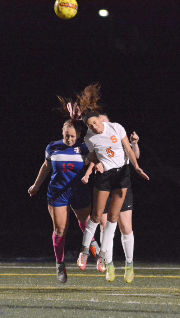
    High school injury reports analyzed by InvestigateWest and Pamplin Media show that girls are twice as likely to get concussions as boys in Oregon. Girls in the 13U age group, pictured above, are the youngest allowed to use headers.
     

    The Concussion Gap: Head injuries in girls soccer are an ‘Unpublicized Epidemic’

    Lee van der Voo, InvestigateWest, photos by David Ball / Pamplin Media Group

     
    When it comes to concussion in sports, all eyes are on football, or so it seems. But it’s not just football that causes a high number of head injuries among young athletes.

    Another culprit? Girls soccer.

    National research has found girls are more likely to suffer a concussion than boys in any sport. In 2017, researchers at Northwestern University generated national headlines when they found concussion rates among young female soccer players were nearly as high as concussion rates for boys playing football — and roughly triple the rate of concussions in boys soccer.

    In Oregon, injury reports from public high schools analyzed by InvestigateWest and Pamplin Media Group mirrored that trend, showing soccer concussions were second to those from football between 2015 and 2017. What’s more, at the schools that included the gender of injured athletes, there were nearly twice as many reports of possible concussions for girls playing soccer than boys in the sport.

    The rate of concussions in girls soccer worries local experts like Jim Chesnutt, a doctor in sports medicine at Oregon Health & Science University, who says those injuries are not widely recognized, even as concussion rates rise for girls playing soccer.

    “In a lot of ways, it’s a growing epidemic for young girls that I think has gone unpublicized,” said Chesnutt, co-director of the Oregon Concussion Awareness and Management Program and a member of the Governor’s Task Force on Traumatic Brain Injury.

    More exposure, more injury

    It’s understandable that much of the youth concussion conversation centers on football, given the physical contact that is visibly — and audibly — evident on every play, as well as the large rosters and the lengthy lists of players who are injured.

    But if you compare girls soccer with football, and only look at the high school participation and injury data, “you’re missing a gigantic part of the picture,” according to Michael Koester, a doctor of sports medicine at the Slocum Center in Eugene. He directs its sports concussion program and serves as the chair of the Sports Medicine Advisory Committee for the National Federation of State High School Associations.

    Koester notes that high school boys play eight to 10 football games per season, and typically play other sports in the off-season.

    Girls, however, play 15 to 20 soccer games in a high school season, but when that season ends, they may play another 80-plus games throughout the winter, spring and summer with club teams, said Koester, who, like Chesnutt, is a medical adviser to the Oregon Schools Activities Association.

    “If we’re looking at injury risk by athletic exposure,” which is one practice or game, a standard in evaluating risk, Koester said, female soccer players probably are playing five if not 10 times more practices and games than football players.

    And Koester doesn’t see the trend ending.

    “The thought used to be that this was all revolving around, ‘Wow! They want to get their kid a scholarship,’ ” he said. “Now it’s kind of gotten to the point where there’s so much single-sport participation that we see kids that are specializing in sport early, just so they’ll be able to make their high school team.”

    Single-sport athletes are more prone to injury in any sport. According to a study by scientists at the University of Wisconsin, high school athletes who specialized in just one sport at an early age were twice as likely to suffer injuries to their lower extremities.

    “We see a lot of overuse injury among girls playing soccer,” Koester said. “We see a lot of ACL injury among girls playing soccer. It’s a well-known problem.”

    Aggressive play

    Another factor is the evolution of sports.

    Angella Bond is an athletic trainer for Tuality Sports Medicine and works on the sidelines with athletes at Hillsboro schools. Anecdotally, she said, all athletes push to be bigger, faster and stronger. Soccer is no exception, nor are girls.

    As athletes develop, they take bigger hits at higher speeds, and competitive games build on their momentum. As competition grows in girls soccer, the sport is trending to be more aggressive, she said.

    “Unfortunately, I think that happens with girls sports,” she said. “Arms fly a little bit more.”

    Chesnutt agreed. “I think over the years, soccer has become more physical,” he said. “And I think the physical contact and the aggressive nature of that physical contact is more associated with concussions.”

    According to the American Academy of Pediatrics, soccer — unlike football, ice hockey and lacrosse — is not a “collision sport.” But it is a “contact sport” because athletes “routinely make contact with each other or inanimate objects.”

    Header balls, though often singled out as a source of concussions, are not necessarily to blame.

    The force created when a soccer ball meets a head can rattle a brain, but data increasingly points to other factors when competitors vie for a ball in the air.

    According to a study by The Research Institute at Nationwide Children’s Hospital, while headers accounted for 27 percent of concussions, it was knocks with other players on aerial play — including head-to-head contact and arms and elbows to the head — and contact with the ground that accounted for 70 percent of those concussions in girls soccer, suggesting aggressive play is a factor in most concussions involving headers.

    Why girls?

    But why are girls more prone to concussions than boys while playing soccer? The prevailing theories focus on their weaker neck-muscle development, weaker body strength (needed to stabilize the neck and head during aerial play), and more frequent contact with the ground. A year ago, a study in the Journal of the American Osteopathic Association found that female high school soccer players took twice as long as male players to recover.

    It’s also possible that girls don’t benefit as much from early treatment. A recent study published by the American Academy of Pediatrics found that girls are five times more likely than boys to stay on the pitch and play through a head injury.

    And the soccer community has been slow to recognize the hard hits its girls are taking. Instead, soccer is at the forefront of the cultural empowerment of girls.

    Local experts concerned about concussion risk note that sports, including girls soccer, have plenty of benefits. Just being physically active is good for kids, and sports like soccer help establish lifelong fitness habits, teach team-building skills, and promote character development and assertiveness.

    “The worry is that the take-home message is that (girls soccer) is healthy and fantastic and nothing can be bad about it,” said Koester, who says an opposite negative message, equally extreme, is more often associated with boys playing football.

    Greater awareness needed

    Concussion education and awareness in girls soccer is paramount, according to local experts such as Chesnutt.

    “I think the way to decrease it is to really analyze how we can modify the amount of body contact that goes on in soccer to limit the dangerous aggressive behavior that is associated with concussion,” he said.

    Unlike youth football, a sport that’s adjusting to new information about concussions all the time, soccer has largely failed to address new information about concussions, Chesnutt said.

    Football, for example, has reduced head-to-head helmet play, limited full-contact practices and games, and zeroed in on the specialty teams with the highest concussion rates.

    “Football has really done, I think, an exceptional job of identifying some areas where there have been some definite higher incidents and some problems,” said Chesnutt, who lectures nationally about youth concussions. “As a group of coaches, leagues, parents and referees, they’ve all looked at it and come up with some solutions that have decreased concussion rates. And I think it’s time for soccer to do the same thing.”

    Read the original article


    Top 10 Volunteer Opportunities in San Diego in 2019

    September 25, 2019, by Mary at greatnonprofits.org

     
    Want to volunteer or intern at a great San Diego nonprofit? Whether you’re new to the city and want to learn about its charities, trying to change up your routine with some local charity work, or just want to volunteer or intern at a neighborhood nonprofit, everyone knows that the best way to find the right place for you is from the people who’ve been there!

    Here’s a list of volunteers’ and interns’ favorite San Diego charities. Every nonprofit on this list has earned an overall score of 4 or greater out of 5 on GreatNonprofits.org. If your favorite San Diego nonprofit or volunteer gig is missing, find it on GreatNonprofits.org, write a positive review, and show your co-volunteers how to start adding reviews and get it on the list!

    Mayan Families
    We just returned after 10 days working with Mayan Families. I, along with my daughter and nephew, have been volunteering with this great nonprofit for the past four years. The focus of our volunteering has been to raise money for the purpose of installing stoves for indigenous families living around Lake Atitlán. The beauty of this particular program, and most of the programs run by Mayan Families, is the direct and immediate impact they have on the recipients. We love the fact that we see where the money we raised is going and that we literally have a hand in helping change the lives of people who truly need the help.

    “We continue to be impressed with Mayan Families’ dedication to its motto to ‘Educate, Feed, Shelter, Feed’ these wonderful people around Lake Atitlán.” –David Kujan

    Sepsis Alliance
    “As a small nonprofit, they do a tremendous job of spreading awareness about sepsis and as a result have reached millions of people to educate them about the signs and symptoms of this condition, albeit with their limited staff and budget.

    “I feel confident in asking others for donations for this organization, as I have seen firsthand that they use their funds very effectively.” –Lynn S1

    Labrador Rescuers
    “Lab Rescue goes over and above to help match the right family with the right lab.

    They have a great foster program that provides information about the traits of the labs to help find the right fit. We can improve our program by increasing the number of people helping to promote intake, fostering, adoptions, and fundraising.” –mobileUser381273

    San Diego Dance Theatre
    “The Dance Fierce program has served as an incredible creative outlet for students from all backgrounds and has united these students through the art of dance. Students who participate in this program are more well-rounded, expressive, and balanced. They pride themselves on their hard work and are more motivated every day through their experiences.” –Mmctighe

    San Diego Brain Injury Foundation
    “I ended up doing one of my internships at SDBIF. Never have I seen so few accomplish so much for so many on so little resources.

    I can only imagine how much more dynamic and influential in helping those with brain injuries, myself included, this organization could be if they had additional funding. The ‘F’ signifying foundation should be changed to ‘Family’—as this organization helps us all to feel this way during very trying times that can last for years.” –Michael Murphy

    College Area Pregnancy Services
    “During the almost 14 years that I volunteered at CAPS as a counselor I witnessed firsthand the impact this place has on every client who comes in. Women from all ages come burdened with fear, confusion, and uncertainty. Volunteers and staff at CAPS are able to provide a safe, nonjudgmental place for these women where they find not only help and resources, but also a caring and personal environment. A comment I most often heard after a counseling appointment was ‘This place is so nice, I felt comfortable and welcomed here.’

    “CAPS will be forever in my heart, and love to tell others about it.” –Ana_39

    The League of Amazing Programmers
    “The League has done an incredible job exposing young people to the vast world of computers in a way that is fun and interactive!

    As a volunteer, I have seen kids develop confidence and problem-solving prowess before my very eyes, all while developing skills they will use for the rest of their lives!” –Mike D3

    Mind Treasures
    “I’ve had the privilege of volunteering with this wonderful organization for many years. Their program is changing the lives of the children one student and school at the time. Children are becoming aware of their hidden potentials and learning how to use these resources in their personal, family, and community finances.” –MT Volunteer 1

    The Seany Foundation
    “The passion that you see from those involved in this foundation is infectious. From the founders, board members, organizers, and volunteers you see an intense commitment to carry on the fight for whom this foundation is in honor of, Sean Robins. The rapidly accelerating success in awareness and donations is a testament to their effectiveness as an organization and their tremendous potential.” –Keenan 27

    Voice of the Bride Ministries
    “Voice of the Bride is a beautiful expression of community love and hard work. I’m constantly amazed at how far they manage to stretch each dollar and how many people they touch—be it by feeding families, helping community, or simply being a force of goodness in an area. They truly love the poor and give to the needy.” –FreckldFlower

    Read the original article online
     

    Serving the Brain Injury Community Since 1983