News

Brain wrapped in rubbberbands to demonstrate neuroplasticity

Does Brain Plasticity Increase After a Head Injury?

Medically reviewed by Nancy Hammond, M.D.
By James Roland for healthline.com on July 28, 2022

 
Brain plasticity, also called neuroplasticity, refers to the brain’s ability to adapt its structure and function in response to changes, such as a head injury or aging. Brain plasticity also involves the formation of new connections between neurons (brain cells).

The brain’s ability to reorganize these features after an injury affects the nature of post-injury recovery.

The severity of the injury goes a long way toward determining how the brain responds. But it’s often possible to boost brain plasticity with interventions and rehabilitation during the healing process.

What is brain plasticity?
Brain plasticity is a term that refers to the brain’s ability to restructure and reconfigure itself in response to change.

Change that can influence the brain comes in several forms. Expected changes include learning, experience, and aging. Unexpected changes include things like stroke and head injury.

Neuroplasticity has long been observed in children. It involves a process called neurogenesis, which is the formation of new neurons in the brain (and elsewhere in the nervous system).

There are two basic types of brain plasticity: structural and functional.

Structural Plasticity
Structural plasticity refers to the way the brain’s physical structure changes in response to learning.

For example, a small 2018 studyTrusted Source showed that healthy adults who participated in balance training twice a week, for 12 weeks, experienced thickening in certain areas of the brain involved in spatial orientation.

A 2016 study examined neuroplasticity in people learning to read Braille. It found that over the course of daily lessons, for 3 weeks, study participants developed increased connectivity in regions of the brain involved in processing sensations like touch.

Functional Plasticity
Functional plasticity refers to the brain’s ability to heal itself after injury. To achieve this, healthy regions of the brain adapt to take over certain functions that the damaged parts of the brain used to perform. This makes functional plasticity especially relevant for people recovering from head injuries.

A 2017 review of studies examining the role of neuroplasticity in stroke recovery found that a stroke can actually trigger neuroplasticity in certain areas. Neuroplasticity plays a role as the brain tries to resume regular functions, like speaking and controlling the movement of limbs.
 
Brain image with new wiring

Can brain plasticity help you heal after a TBI?

 
A traumatic brain injury (TBI) refers to changes in brain function or brain health caused by an external force, such as a serious blow to the head.

The Centers for Disease Control and Prevention (CDC)Trusted Source reports that there were more than 220,000 TBI-related hospitalizations in 2019 and more than 64,000 TBI-related deaths the following year.

A TBI differs from a nontraumatic brain injury, also known as an acquired brain injury. Acquired brain injuries are those caused by internal factors, such as a stroke, which can damage brain tissue and affect muscle control, speech, cognition, and other functions.

When spontaneous brain plasticity doesn’t occur, it’s sometimes possible to boost neuroplasticity artificially.

 
A 2020 review of neuroplasticity therapies to treat stroke survivors suggests that approaches such as brain stimulation therapy and virtual reality might help enhance brain plasticity. It may also be possible to transfer nerves from healthy parts of the brain to injured parts.

Similarly, a 2017 review of studies on cognitive rehabilitation following TBI, suggests that memory and other thinking skills may be recovered to some degree with the help of cognitive rehabilitation. The studies showed how cognitive rehabilitation helped to modify damaged neural connections and various brain functions.

Does a brain injury increase neuroplasticity?

Because different regions of the brain are responsible for different functions, the location and severity of an injury determine which functions are affected and to what degree.

For example, certain areas of the brain are responsible for your ability to move certain parts of the body, like your left arm or your right foot.

This is where brain plasticity can help you heal after a brain injury. Just as exercise and learning can enhance brain structure and function, the body’s natural healing and recovery process after an injury can also increase neuroplasticity.

When neurons die due to injury, the brain naturally responds within a few days by developing new neural networks and recruiting various types of cells to take the place of those damaged or killed in the injury.

The extent to which neuroplasticity occurs depends on an individual’s age, the location of the injury, and other factors.

Does age matter after brain injury?

Whether it’s a brain injury or a broken wrist, being younger is always an advantage when it comes to recovery.

A 2008 studyTrusted Source of TBI survivors noted that disability scores following a TBI tended to be significantly better among younger TBI survivors compared with older individuals, even when those older survivors had less severe injuries. And the younger patients improved more in the first 5 years after the injury.

A 2019 report notes that because age affects neuroplasticity, the need for more strategies and therapies to compensate for age-related changes should be a higher priority in the face of an aging population.

Can you see brain plasticity on an MRI?

One of the most useful tools in diagnosing the impact of a TBI, stroke, or other injury or illness affecting the brain is magnetic resonance imaging (MRI).

An MRI can detect many changes in brain structure and function. Current technology is far from perfect, but it’s continuing to improve.

A 2021 articleTrusted Source suggests that advanced MRI techniques are helping doctors develop a more accurate picture of mild TBIs. This may help improve the treatment and understanding of mild TBIs in the future.

A newer type of MRI, called functional MRI (fMRI), can help doctors observe brain activity, not just brain structure. This may be particularly helpful in studying brain damage and recovery.

A 2017 studyTrusted Source of neuroimaging after TBI notes that fMRI can detect changes in thinking skills, emotions, and the course of neuroplasticity after an injury to the brain. The study says that fMRI is a helpful tool in assessing the damage caused by TBI and tracking brain changes during recovery.

But fMRI, the study says, will need to be accompanied by other data if it’s going to inform treatment decisions. This includes information gathered during cognitive-behavioral evaluations and other assessments.
Image Neurons reconnecting

How long does it take to heal after a TBI?

 
The time necessary to heal from a TBI can vary considerably from one person to the next. This is based mostly on the seriousness of the injury, as well as its location, the age of the individual, and that person’s overall physical and mental health.

A full recovery from a mild TBI can be expected in about 3 months. People with a moderate TBI will take longer to heal and will typically need cognitive rehabilitation, physical therapy, and other interventions.

Predicting the degree and length of recovery from a severe TBI is very difficult, and should be done on a case-by-case basis.

Takeaway

Brain plasticity after a head injury is when brain functions thought to be lost due to damage begin to be adopted by other, healthy brain tissue.

While not all functions can be reorganized or reestablished completely, the brain’s remarkable adaptability can often help people who had a stroke, traumatic brain injury, or other harmful events recover some function.

Brain plasticity can be encouraged through cognitive therapy, physical therapy, and other treatments.

CLICK HERE to read the original article
 

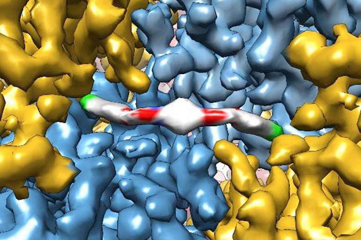
ISRIB molecule—image by the Adam Frost lab at UCSF

Drug Reverses Age-Related Mental Decline Within Days, Suggesting Lost Cognitive Ability is Not Permanent

By Good News Network, December 27, 2020

 
Just a few doses of an experimental drug that reboots protein production in cells can reverse age-related declines in memory and mental flexibility in mice, according to a new study by UC San Francisco scientists.

The drug, called ISRIB, has already been shown in laboratory studies to restore memory function months after traumatic brain injury (TBI), reverse cognitive impairments in Down Syndrome, prevent noise-related hearing loss, fight certain types of prostate cancer, and even enhance cognition in healthy animals.

In the new study, published Dec. 1 in the open-access journal eLife, researchers showed rapid restoration of youthful cognitive abilities in aged mice, accompanied by a rejuvenation of brain and immune cells that could help explain improvements in brain function—and with no side effects observed.

“ISRIB’s extremely rapid effects show for the first time that a significant component of age-related cognitive losses may be caused by a kind of reversible physiological “blockage” rather than more permanent degradation,” said Susanna Rosi, PhD, Lewis and Ruth Cozen Chair II and professor in the departments of Neurological Surgery and of Physical Therapy and Rehabilitation Science.

“The data suggest that the aged brain has not permanently lost essential cognitive capacities, as was commonly assumed, but rather that these cognitive resources are still there but have been somehow blocked, trapped by a vicious cycle of cellular stress,” added Peter Walter, PhD, a professor in the UCSF Department of Biochemistry and Biophysics and a Howard Hughes Medical Institute investigator. “Our work with ISRIB demonstrates a way to break that cycle and restore cognitive abilities that had become walled off over time.”

Rebooting cellular protein production holds key to aging

Walter has won numerous scientific awards, including the Breakthrough, Lasker and Shaw prizes, for his decades-long studies of cellular stress responses. ISRIB, discovered in 2013 in Walter’s lab, works by rebooting cells’ protein production machinery after it gets throttled by one of these stress responses—a cellular quality control mechanism called the integrated stress response (ISR; ISRIB stands for ISR InhiBitor).

The ISR normally detects problems with protein production in a cell—a potential sign of viral infection or cancer-promoting gene mutations—and responds by putting the brakes on cell’s protein-synthesis machinery. This safety mechanism is critical for weeding out misbehaving cells, but if stuck in the ‘on’ position in a tissue like the brain, it can lead to serious problems, as cells lose the ability to perform their normal activities, according to Walter and colleagues.

In particular, their recent animal studies have implicated chronic ISR activation in the persistent cognitive and behavioral deficits seen in patients after TBI, by showing that, in mice, brief ISRIB treatment can reboot the ISR and restore normal brain function almost overnight.

The cognitive deficits in TBI patients are often likened to premature aging, which led Rosi and Walter to wonder if the ISR could also underlie purely age-related cognitive decline. Aging is well known to compromise cellular protein production across the body, as life’s many insults pile up and stressors like chronic inflammation wear away at cells, potentially leading to widespread activation of the ISR.

“We’ve seen how ISRIB restores cognition in animals with traumatic brain injury, which in many ways is like a sped-up version of age-related cognitive decline,” said Rosi, who is director of neurocognitive research in the UCSF Brain and Spinal Injury Center and a member of the UCSF Weill Institute for Neurosciences. “It may seem like a crazy idea, but asking whether the drug could reverse symptoms of aging itself was just a logical next step.”

Signature effects of aging disappeared literally overnight

In the new study, researchers led by Rosi lab postdoc Karen Krukowski, PhD, trained aged animals to escape from a watery maze by finding a hidden platform, a task that is typically hard for older animals to learn. But animals who received small daily doses of ISRIB during the three-day training process were able to accomplish the task as well as youthful mice—and much better than animals of the same age who didn’t receive the drug.

The researchers then tested how long this cognitive rejuvenation lasted and whether it could generalize to other cognitive skills. Several weeks after the initial ISRIB treatment, they trained the same mice to find their way out of a maze whose exit changed daily—a test of mental flexibility for aged mice who, like humans, tend to get increasingly stuck in their ways. The mice who had received brief ISRIB treatment three weeks before still performed at youthful levels, while untreated mice continued to struggle.

To understand how ISRIB might be improving brain function, the researchers studied the activity and anatomy of cells in the hippocampus, a brain region with a key role in learning and memory, just one day after giving animals a single dose of ISRIB. They found that common signatures of neuronal aging disappeared literally overnight: neurons’ electrical activity became more sprightly and responsive to stimulation, and cells showed more robust connectivity with cells around them while also showing an ability to form stable connections with one another usually only seen in younger mice.

The researchers are continuing to study exactly how the ISR disrupts cognition in aging and other conditions and to understand how long ISRIB’s cognitive benefits may last. Among other puzzles raised by the new findings is the discovery that ISRIB also alters the function of the immune system’s T cells, which also are prone to age-related dysfunction. The findings suggest another path by which the drug could be improving cognition in aged animals, and could have implications for diseases from Alzheimer’s to diabetes that have been linked to heightened inflammation caused by an aging immune system.

“This was very exciting to me because we know that aging has a profound and persistent effect on T cells and that these changes can affect brain function in the hippocampus,” said Rosi. “At the moment, this is just an interesting observation, but it gives us a very exciting set of biological puzzles to solve.”

Success shows the ‘serendipity’ of basic research

Rosi and Walter were introduced by neuroscientist Regis Kelly, PhD, executive director of the University of California’s QB3 biotech innovation hub, following Walter’s 2013 study showing that the drug seemed to instantly enhance cognitive abilities in healthy mice. To Rosi, the results from that study implied some walled-off cognitive potential in the brain that the molecule was somehow unlocking, and she wondered if this extra cognitive boost might benefit patients with neurological damage from traumatic brain injury.

The labs joined forces to study the question in mice, and were astounded by what they found. ISRIB didn’t just make up for some of the cognitive deficits in mice with traumatic brain injury—it erased them. “This had never been seen before,” Rosi said. “The mantra in the field was that brain damage is permanent—irreversible. How could a single treatment with a small molecule make them disappear overnight?”

Further studies demonstrated that neurons throughout the brains of animals with traumatic brain injury are thoroughly jammed up by the ISR. Using ISRIB to release those brakes lets brain cells immediately get back to their normal business. More recently, studies in animals with very mild repetitive brain injury—akin to pro athletes who experience many mild concussions over many years—showed that ISRIB could reverse increased risk-taking behavior associated with damage to self-control circuits in the frontal cortex.

“It’s not often that you find a drug candidate that shows so much potential and promise,” Walter says, calling it “just amazing”.

No side effects

One might think that interfering with the ISR, a critical cellular safety mechanism, would be sure to have serious side effects, but so far in all their studies, the researchers have observed none. This is likely due to two factors. First, it takes just a few doses of ISRIB to reset unhealthy, chronic ISR activation back to a healthier state. Second, ISRIB has virtually no effect when applied to cells actively employing the ISR in its most powerful form—against an aggressive viral infection, for example.

ISRIB has been licensed by Calico, a South San Francisco, Calif. company exploring the biology of aging, and the idea of targeting the ISR to treat disease has been picked up by many other pharmaceutical companies, Walter says.

“It almost seems too good to be true, but with ISRIB we seem to have hit a sweet spot for manipulating the ISR with an ideal therapeutic window,” Walter said.

Get more links to background studies from original article from UCSF News.
 

CLICK HERE to read the original article


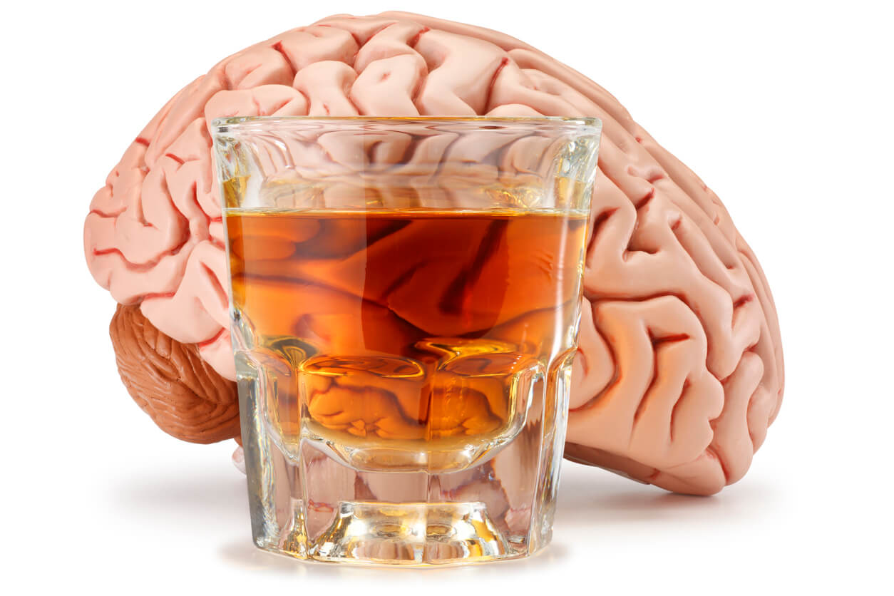
Alcohol and Brain Injury

After a TBI or Stroke the brain can become more sensitive to the effects of alcohol. This can cause cognitive problems that impact memory, mobility, and speech. It can also cause someone to feel fatigued and unwell. What can be expected after your recovery?


By Fred at Spindpals.com, July 18, 2020


Whether we’re on holiday abroad or enjoying the Christmas festivities, an alcoholic drink tends to not be too far from reach for many of us.

But after a brain injury, the body’s tolerance to alcohol is greatly reduced, and many survivors find that they are no longer able to enjoy alcohol in the same way as they did before their injury. The reduced tolerance to alcohol means that many effects of brain injury are exacerbated after drinking, such as memory problems, mobility issues, speech and fatigue.

Remember you should always discuss with your medical practitioner your particular condition to understand what the impact would be on yourself. Never take alcohol without their approval and guidance.

AUTHOR: “It is clear that there is an uneasy relationship between alcohol and brain injury. Survivors are often faced with the challenge of balancing a desire to enjoy the social life they had before they sustained their injury with the acceptance that alcohol now affects them in a different way.”

We asked brain injury survivors to tell us about how their relationship with alcohol has changed.

For some, the enjoyment of drinking is simply outweighed by the effects caused.

“I don’t drink anymore,” said Louise Fry. “I couldn’t drink to start with because of meds, but now? It just hits me too hard.”

Janet Creamer agreed: “Drinking is now a no-no. Just one alcoholic drink does awful things to my brain. It feels like I’ve drunk way too much and I get that spaced out feeling.”

Others, like Giles Philip Hudson, have found that being advised by doctors to no longer drink has actually been a blessing in disguise.

AUTHOR: “After sustaining my brain injury and spending over four months in hospital, doctors advised me not to drink alcohol. During this time I found I no longer needed to drink alcohol to make me feel good or enjoy myself. I certainly don’t need the headaches it causes.”
 

Enjoy a drink at home with family and friends

Naturally, many people want to continue to be able to enjoy a drink every now and then, particularly at social gatherings. But what if going to the bar is too daunting a prospect?

Home drinking is increasingly popular For some, staying in allows them to enjoy a drink without some of the challenges of being in a busy, crowded and noisy pub or bar.

“Since my disability I do not feel comfortable going into a bar as I may find it hard to use the restrooms,” said one member of the community, “so my drinking is done in my home.”

Patricia Nugent on Facebook agreed: “We tend to drink at home so it is easier and less stressful to moderate intake,” she said.

If you are choosing to drink at home, it’s important you monitor your intake carefully.
Here’s some useful advice for home drinkers:

    1. Keep track of how many units you’re consuming
    2. Use smaller glasses
    3. Use proper spirit measures to avoid inadvertently pouring yourself a double or triple measure
    4. Eat as you drink
    5. Invest in a good bottle stop to make that bottle of wine last longer

Out and about

For others, however, a good night out is still a must! If that’s the case, then planning ahead can be the key to the success of the evening.

“I don’t go out much, once every two months,” said Michelle Richardson. “But it’s lovely to have some drinks and let my hair down and forget how challenging recovery is for a while.

AUTHOR: “I do have to prepare for a night out by having an afternoon snooze.”

If you do want to enjoy a night out on the town with friends, here are some more top tips:

    ▪ Don’t drink on an empty stomach and check your medication allows you to drink
    ▪ Make sure your friends know about your brain injury, lowered alcohol tolerance levels, and any other issues such as an intolerance to noise
    ▪ Drink water between alcoholic drinks and avoid getting into rounds

Alcohol-free alternatives

Of course, not drinking alcohol doesn’t mean you can’t still go out to pubs and bars.

“My husband has been told he can’t drink alcohol,” said Amanda Hopkins. “So, as he is a real ale drinker, we made a pact to still go to country location but to just check out ‘alcohol-free’ ales and to become connoisseurs of the growing ‘alcohol-free’ ranges that are now appearing from many microbreweries.

“It won’t be quite the same but we hope it will be a bit of fun tasting them.”

AUTHOR: Kathy M agreed: “I sometimes have a non-alcoholic beer shandy so I feel like I am having a pint and I’ve discovered things like elderflower cordial with soda. There’s nothing wrong with ordering a fancy coffee or mocktail either.”

Drinking alcohol after Stroke

    ▪ Drinking too much alcohol contributes to a number of risk factors for stroke, including high blood pressure.
    ▪ Alcohol can interfere with the medicine you take to reduce stroke risk.
    ▪ Your doctor can advise when it is safe for you to start drinking alcohol again and how much alcohol it is safe for you to drink.
    ▪ Healthy men and women should have no more than two standard drinks a day, and no more than four standard drinks on any one occasion.

Alcohol and stroke risk

Drinking too much alcohol contributes to a number of risk factors for stroke. If you have already had a stroke or transient ischaemic attack (TIA), you can help reduce your risk by only drinking a safe amount.

High blood pressure is the biggest risk factor for stroke, and drinking too much raises your blood pressure. Atrial fibrillation, which is a type of irregular heartbeat, can be triggered by too much alcohol.

Diabetes and being overweight also increase your risk of having a stroke. Both of are linked to alcohol consumption.

Alcoholic drinks are also high in calories with little nutritional value. Reducing the amount you drink will support you to maintain a healthy weight.

Hemorrhagic stroke and alcohol

A hemorrhagic stroke is caused by a break in the wall of a blood vessel in the brain. If you have had a hemorrhagic stroke, you must not drink alcohol for at least three weeks after your stroke. Ask your doctor when it is safe to start drinking alcohol again.

Drinking alcohol and your medication

Alcohol could interfere with the medicine you take particularly, blood-thinning medicine such as Warfarin. Discuss with your doctor about whether it is safe to drink alcohol while taking any medicines.

Consuming alcohol safely if your doctor clears you to

The Guidelines for Alcohol Consumption gives advice about safe amounts of alcohol.

AUTHOR: Remember, the Guidelines are for healthy people. Talk to your doctor about whether it is safe for you to drink at all, and whether the amounts in the Guidelines are safe for you.

The Guidelines state that healthy men and women should have no more than two standard drinks on any day and if you go out, no more than four standard drinks on any one occasion.

    ▪ For spirits with 40 % ABV, a standard drink is 30 mls (1.5 fl oz)
    ▪ A 285 ml (10 fl oz) glass of 3.5% ABV beer is about 1 standard drink.
    ▪ 100 ml (3.5 fl oz) of wine or champagne is approximately one standard drink, however this varies between types. Keep in mind most glasses of wine served in restaurants and bars are more than 100 ml (3.5 fl oz).
    ▪ Always check the label on the bottle to find out how many standard drinks you are having.
    If you find you are tempted to go over your safe limits learn strategies to help you keep to them.

Strategies to reduce your drinking

Write down how many drinks you have to see how much and how often you drink.
If you find that you are drinking more than is safe, try these tips:

    ▪ Drink water when you are thirsty rather than alcohol.
    ▪ Sip your drink slowly. Put down the glass after each mouthful.
    ▪ At social occasions, make every second drink a non-alcoholic beverage. Choose something like a sparkling water rather than a sugary drink.
    ▪ Try low-alcohol alternatives such as light beer.
    ▪ Opt out of ‘shouts’. Drink at your own pace. If you cannot avoid buying a round, get yourself a non-alcoholic drink.
    ▪ Avoid salty snacks such as potato chips or peanuts. These make you thirsty and more inclined to drink quickly.
    ▪ Set goals such as not drinking alone and have at least two days without alcohol each week.
    ▪ Do not drink on an empty stomach. A full stomach slows the absorption of alcohol

Brain Injury affects different people in different ways. There is no one size fits all. For some, their relationship with alcohol will be over. For others, a moderate consumption can be tolerated. Complete your recovery and then discuss with your medical practitioner your options. Do not make any decisions without consulting them first.

This post is shared from the Stroke Association AU and Headway UK websites
 
CLICK HERE to read the original article


Ayn al Asad Air Base in western Iraq after an Iranian missile attack on Jan. 8. The number of service members experiencing symptoms associated with brain injuries has since topped 100. Photo Credit…Sergey Ponomarev for The New York Times


Brain Injuries Are Common in Battle.
The Military Has No Reliable Test for Them.

Traumatic brain injury is a signature wound of the wars in Iraq and Afghanistan. But the military still has no objective way of diagnosing it in the field.

By Dave Philipps and Thomas Gibbons-Neff for nytimes.com, February 15, 2020

 
U.S. troops at Ayn al Asad Air Base in western Iraq hunkered down in concrete bunkers last month as Iranian missile strikes rocked the runway, destroying guard towers, hangars and buildings used to fly drones.
When the dust settled, President Trump and military officials declared that no one had been killed or wounded during the attack. That would soon change.

A week after the blast, Defense Department officials acknowledged that 11 service members had tested positive for traumatic brain injury, or TBI, and had been evacuated to Kuwait and Germany for more screening. Two weeks after the blast, the Pentagon announced that 34 service members were experiencing symptoms associated with brain injuries, and that an additional seven had been evacuated. By the end of January the number of potential brain injuries had climbed to 50. This week it grew to 109.

The Defense Department says the numbers are driven by an abundance of caution. It noted that 70 percent of those who tested positive for a TBI had since returned to duty. But experts in the brain injury field said the delayed response and confusion were primarily caused by a problem both the military and civilian world have struggled with for more than a decade: There is no reliable way to determine who has a brain injury and who does not.

Top military leaders have for years called traumatic brain injury one of the signature wounds of the wars in Iraq and Afghanistan; at the height of the Iraq war in 2008, they started pouring hundreds of millions of dollars into research on detection and treatment. But the military still has no objective tool for diagnosing brain injury in the field. Instead, medical personnel continue to use a paper questionnaire that relies on answers from patients — patients who may have reasons to hide or exaggerate symptoms, or who may be too shaken to answer questions accurately.

The military has long struggled with how to address so-called invisible war wounds, including traumatic brain injury and post-traumatic stress disorder. Despite big investments in research that have yielded advances in the laboratory, troops on the ground are still being assessed with the same blunt tools that have been in use for generations.

The problem is not unique to the military. Civilian doctors struggle to accurately assess brain injuries, and still rely on a process that grades the severity of a head injury in part by asking patients a series of questions: Did they black out? Do they have memory problems or dizziness? Are they experiencing irritability or difficulty concentrating?

“It’s bad, bad, bad. You would never diagnose a heart attack or even a broken bone that way,” said Dr. Jeff Bazarian a professor of emergency medicine at the University of Rochester Medical Center. “And yet we are doing it for an injury to the most complex organ in the body. Here’s how crazy it gets: You are relying on people to report what happened. But the part of the brain most often affected by a traumatic brain injury is memory. We get a lot of false positives and false negatives.”

Without a good diagnosis, he said, doctors often don’t know whether a patient has a minor concussion that might require a day’s rest, or a life-threatening brain bleed, let alone potential long-term effects like depression and personality disorder.

At Ayn al Asad, personnel used the same paper questionnaires that field medics used in remote infantry platoons in 2010. Aaron Hepps, who was a Navy corpsman in a Marines infantry company in Afghanistan at that time, said it did not work well then for lesser cases, and the injuries of many Marines may have been missed. During and after his deployment, he counted brain injuries in roughly 350 Marines — about a third of the battalion.

After the January missile attack, Maj. Robert Hales, one of the top medical providers at the air base, said that the initial tests were “a good start,” but that it took numerous screenings and awareness among the troops to realize that repeated exposure to blast waves during the hourlong missile strikes had affected dozens.

Traumatic brain injuries are among the most common injuries of the wars in Iraq and Afghanistan, in part because armor to protect from bullet and shrapnel wounds has gotten better, but they offer little protection from the shock waves of explosions. More than 350,000 brain injuries have been reported in the military since 2001.

The concrete bunkers scattered around bases like Ain al Assad protect from flying shrapnel and debris, but the small quarters can amplify shock waves and lead to head trauma.

The blasts on Jan. 8, one military official said, were hundreds of times more powerful than the rocket and mortar attacks regularly aimed at U.S. bases, causing at least one concrete wall to collapse atop a bunker with people inside.

Capt. Geoff Hansen was in a Humvee at Ayn al Asad when the first missile hit, blowing open a door. Then a second missile hit.

“That kind of blew me back in,” he said. “Blew debris in my face so I went and sat back down a little confused.”

A tangle of factors make diagnosing head injuries in the military particularly tricky, experts say. Some troops try to hide symptoms so they can stay on duty, or avoid being perceived as weak. Others may play up or even invent symptoms that can make them eligible for the Purple Heart medal or valuable veteran’s education and medical benefits.

And sometimes commanders suspect troops with legitimate injuries of malingering and force them to return to duty. Pentagon officials said privately this week that some of the injuries from the Jan. 8 incident had probably been exaggerated. Mr. Trump seemed to dismiss the injuries at a news conference in Davos, Switzerland, last month. “I heard they had headaches,” he said. “I don’t consider them very serious injuries relative to other injuries I have seen.”

In the early years of the war in Iraq, troops with concussions were often given little medical treatment and were not eligible for the Purple Heart. It was only after clearly wounded troops began complaining of poor treatment that Congress got involved and military leaders began pressing for better diagnostic technology.

Damir Janigro, who directed cerebrovascular research at the Cleveland Clinic for more than a decade, said relying on the questionnaire makes accurate diagnosing extremely difficult.

“You have the problem of the cheaters, and the problem of the ones who don’t want to be counted,” he said. “But you have a third problem, which is that even if people are being completely honest, you still don’t know who is really injured.”

In civilian emergency rooms, the uncertainty leads doctors to approve unnecessary CT scans, which can detect bleeding and other damage to the brain, but are expensive and expose patients to radiation. At the same time doctors miss other patients who may need care. In a war zone, bad calls can endanger lives, as troops are either needlessly airlifted or kept in the field when they cannot think straight.

Mr. Janigro is at work on a possible solution. He and his team have developed a test that uses proteins found in a patient’s saliva to diagnose brain injuries. Other groups are developing a blood test.

Both tests work on a similar principle. When the brain is hit by a blast wave or a blow to the head, brain cells are stretched and damaged. Those cells then dispose of the damaged parts, which are composed of distinctive proteins. Abnormal levels of those proteins are dumped into the bloodstream, where for several hours they can be detected in both the blood and saliva. Both tests, and another test being developed that measures electrical activity in the brain, were funded in part by federal grants, and have shown strong results in clinical trials. Researchers say they could be approved for use by the F.D.A. in the next few years.

The saliva test being developed by Mr. Janigro will look a bit like an over-the-counter pregnancy test. Patients with suspected brain injuries would put sensors in their mouths, and within minutes get a message that says that their brain protein levels are normal, or that they should see a doctor.

But the new generation of testing tools may fall short, said Dr. Gerald Grant, a professor of neurosurgery at Stanford University and a former Air Force lieutenant colonel who frequently treated head injuries while deployed to Iraq in 2005.

Even sophisticated devices had trouble picking up injuries from roadside bombs, he said.

“You’d get kids coming in with blast injuries,” he said, “and they clearly had symptoms, but the CT scans would be negative.”

He was part of an earlier effort to find a definitive blood test, which he said in an interview was “the holy grail.” But progress was slow. The grail was never found, he said, and the tests currently being developed are helpful for triaging cases, but too vague to be revolutionary.

“Battlefield injuries are complex,” he said. “We still haven’t found the magic biomarker.”

CLICK HERE to go to the original article


New Rules to Protect Your Kid’s Noggin

May 25, 2019, Parents Magazine

 
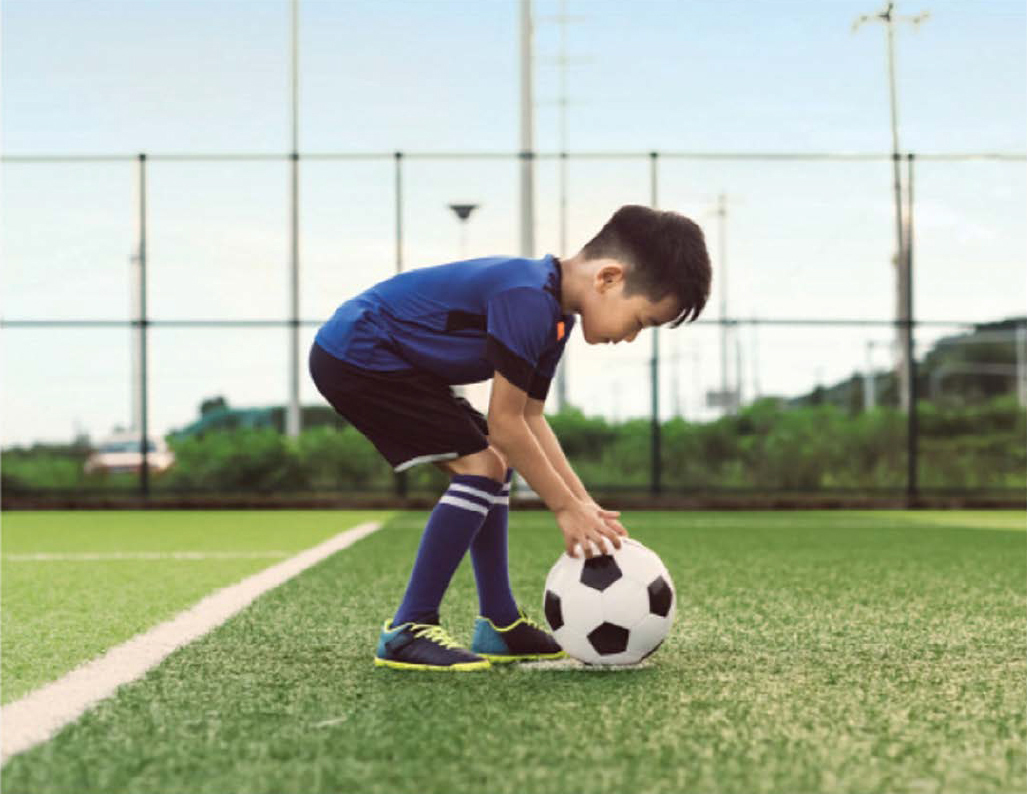
Children bonk their head all the time when they’re wrestling with siblings, playing soccer, and just being clumsy-and it’s easy to worry that a bump could turn into something bigger. After all, more than 800,000 kids in the U.S. get a concussion every year. For the first time, the Centers for Disease Control and Prevention has released specific “return to learn” and “return to play” guidelines for head injuries, based on 25 years of research. One doctor shares the big takeaways.

ALWAYS take any injury beyond a light head bump seiously. A concussion occurs when a bump, blow, or jolt to the head or a hit to the body makes the brain bounce or twist in the skull. This creates chemical changes and can sometimes damage brain cells. “If your child complains of a headache or dizziness, is nauseous or vomiting, appears dazed, or sleeps more or less than usual, it’s time to get a doctor’s evaluation,” says Dennis Cardone, D.O., associate professor of orthopedic surgery and pediatrics and co-director of the NYU Langone Concussion Center. Even toddlers can get a concussion from a tumble, so look for changes in their behavior such as not wanting to nurse or eat or losing interest in toys.

If diagnosed with a concussion, your child will need menlal rest, says Dr. Cardone. That means taking a break from all activities for two to three days, and after that, starting with light aerobic activity. He may need to attend school for only half the day or do little to no homework (he won’t mind this rule!). However, he shouldn’t return to any sports or strenuous activities that have a high risk of falling or contact (think: field hockey, gymnastics, climbing a tree) until he’s been cleared by his doctor, which should be within a few weeks.

Download the original article PDF
 

By Alyssa Navarro, Tech Times (August 23, 2016) — Federal health regulators in the United States approved on Monday the use of two new computer softwares as cognitive screening tests for traumatic head injury patients.

Known as ImPACT or the Immediate Post-Concussion Assessment and Cognitive Testing (ImPACT), the new testing device, as well as a similar test designed for children, can be used by doctors to evaluate signs and symptoms of head injuries that could indicate concussion.

ImPACT is designed for patients aged 12 to 59 years old, while ImPACT Pediatric is intended for children aged 5 to 11 years old, officials said. Licensed health care professionals are the only ones allowed to perform the analysis and interpret the results.

The software can be accessed easily because it runs on both desktop computers and laptops, according to the U.S. Food and Drug Administration (FDA). Both tests the first ever devices permitted by the FDA to assess cognitive function after experiencing a possible concussion. They are designed to be part of medical evaluations in hospitals.

Although ImPACT and ImPACT Pediatric will definitely be useful for doctors, both tests are not meant to diagnose concussions or determine treatments that are appropriate for such cases, the FDA said.

Instead, both devices are only designed to test cognitive skills such as reaction time, memory and word recognition. All of these can be impacted by head injuries. Afterwards, the results are compared to a patient’s pre-injury baseline scores or an age-matched control database, the FDA said.

Dr. Carlos Peña, director of the neurological and physical medicine division at the Center for Devices and Radiological Health, acknowledges that the two testing devices can provide useful information that can aid doctors in the evaluation of people who are experiencing potential signs of concussion.

However, Peña says that clinicians should not completely depend on the tests alone to rule out concussion or to decide whether a player with a head injury should return to a game.

Statistics from the Centers for Disease Control and Prevention (CDC) reveal that traumatic brain injuries are responsible for more than 2 million visits to the emergency room in the country annually. Traumatic brain injuries also account for more than 50,000 deaths in America every year.

Cases of head injury among kids have been increasing. In May, a CDC report showed that from January 2001 to December 2013, approximately 214,883 children aged 14 years old and below were brought to emergency departments due to head injuries.

Read the original article
 

When a person has a stroke, blood flow to the brain is interrupted, causing brain cells to die within minutes due to lack of oxygen. In some cases, this can result in paralysis, speech and language problems, vision problems, and memory loss. But in a new study, researchers have shown that stem cell therapy increases nerve cell production in mice with brain damage due to stroke.

by Marie Ellis, MedicalNewsToday.com (August 22, 2016) — The researchers – led by Berislav Zlokovic, M.D., Ph.D., from the University of Southern California (USC) – publish their findings in the journal Nature Medicine.

According to the Centers for Disease Control and Prevention (CDC), stroke is the fifth leading cause of death in the United States and is also a major cause of disability in adults.

The effects of a stroke depend on the location of the blockage and how much brain tissue is involved, but a stroke on one side of the brain will result in neurological effects on the opposite side of the body.

For example, a stroke on the right side of the brain could produce paralysis on the left side of the body, and vice versa.

A stroke in the brain stem can affect both sides of the body and could leave the patient in a so-called locked-in state, where the patient is unable to speak or move the body below the neck.

Given that about 800,000 people in the U.S. have a stroke each year, the researchers of this latest study wanted to investigate potential therapies.

Therapy is a combination of two methods

The researchers say their therapy is a combination of two methods. One involves surgically grafting human neural stem cells onto the damaged area, where they are able to mature into neurons and other brain cells.

The other therapy uses a compound called 3K3A-APC, which has been shown to help neural stem cells that have been grown in a petri dish grow into neurons. But the researchers say it was not clear what effect the molecule – called activated protein-C (APC) – would have on live animals.

As such, the team used mice for their experiment, and they found that a month after inducing stroke-like brain damage in the mice, those that had received both the stem cells and 3K3A-APC performed much better on motor and sensory function tests, compared with mice that received only one of the treatments or neither.

The researchers also observed that the mice given 3K3A-APC had more stem cells survive and mature into neurons.

But how did the researchers induce stroke-like brain damage in the mice? They disrupted blood flow to a specific brain area.

Then, 1 week later, which is the mouse equivalent of several months in humans, the researchers inserted the stem cells next to the dead tissue and administered either a placebo or 3K3A-APC.

“When you give these mice 3K3A-APC, it works much better than stem cells alone,” says Dr. Zlokovic. “We showed that 3K3A-APC helps the cells convert into neurons and make structural and functional connections with the host’s nervous system.”

‘No one in the stroke field has ever shown this’

The researchers also looked at the connections between the neurons that grew from the stem cells in the damaged brain region and nerve cells in the primary motor cortex.

The team found that the mice given the stem cells and 3K3A-APC had more neuronal connections – synapses – that linked those areas, compared with the mice given the placebo.

Then, when the researchers stimulated the mice’s paws with a vibration, the neurons that grew from the stem cells exhibited a stronger response in the mice that were treated.

“That means the transplanted cells are being functionally integrated into the host’s brain after treatment with 3K3A-APC. No one in the stroke field has ever shown this, so I believe this is going to be the gold standard for future studies.” ~Dr. Berislav Zlokovic

Following on from this study, the researchers want to pursue another phase II clinical trial to examine whether the treatment combination can encourage the growth of new neurons in human stroke patients to improve function.

They say that if that trial is successful, it could be possible to test the therapy’s effects on other conditions, including spinal cord injuries.

“This USC-led animal study could pave the way for a potential breakthrough in how we treat people who have experienced a stroke,” says Jim Koenig, Ph.D., program director at the National Institute of Health’s National Institute of Neurological Disorders and Stroke (NINDS), who funded the study.

“If the therapy works in humans,” he adds, “it could markedly accelerate the recovery of these patients.”

Read the original article

UCSF Researchers Advocate Prioritizing Teens for Education and Prevention

by Scott Maier (August 17, 2016) — The number of Americans diagnosed with concussions is growing, most significantly in adolescents, according to researchers at UC San Francisco. They recommend that adolescents be prioritized for ongoing work in concussion education, diagnosis, treatment and prevention.

The findings appear online August 16, 2016, in the Orthopaedic Journal of Sports Medicine.

“Our study evaluated a large cross-section of the U.S. population,” said lead author Alan Zhang, MD, UCSF Health orthopaedic surgeon. “We were surprised to see that the increase in concussion cases over the past few years mainly were from adolescent patients aged 10 to 19.”

Concussions are a form of mild traumatic brain injury resulting in transient functional and biochemical changes in the brain. They can lead to time lost from sports, work and school, as well as significant medical costs.

Though symptoms resolve in most concussion patients within weeks, some patients’ symptoms last for months, including depression, headache, dizziness and fogginess. Neuroimaging and neuropathological studies also suggest there may be chronic structural abnormalities in the brain following multiple concussions.

Recent studies have shown an increase in traumatic brain injuries diagnosed in many U.S. emergency departments. Smaller cohort studies of pediatric and high school athletes also have indicated a rise in concussions for certain sports, such as football and girls’ soccer. However, this is the first study to assess trends in concussion diagnoses across the general U.S. population in various age groups.

In this study, Zhang and his colleagues evaluated the health records of 8,828,248 members of Humana Inc., a large private payer insurance group. Patients under age 65 who were diagnosed with a concussion between 2007-2014 were categorized by year of diagnosis, age group, sex, concussion classification, and health care setting of diagnosis (emergency department or physician’s office).

Overall, 43,884 patients were diagnosed with a concussion, with 55 percent being male. The highest incidence was in the 15-19 age group at 16.5 concussions per 1,000 patients, followed by ages 10-14 at 10.5, 20-24 at 5.2 and 5-9 at 3.5.

The study found that 56 percent of concussions were diagnosed in the emergency department, 29 percent in a physician’s office, and the remainder in urgent care or inpatient settings. As such, outpatient clinicians should have the same confidence and competence to manage concussion cases as emergency physicians, Zhang said.

A 60 percent increase in concussions occurred from 2007 to 2014 (3,529 to 8,217), with the largest growth in ages 10-14 at 143 percent and 15-19 at 87 percent. Based on classification, 29 percent of concussions were associated with some loss of consciousness.

A possible explanation for the significant number of adolescent concussions is increased participation in sports, said Zhang, MD, who is also assistant professor of orthopaedic surgery at UCSF. It also may be reflective of an improved awareness for the injury by patients, parents, coaches, sports medical staff and treating physicians.

For example, the U.S. Centers for Disease Control and Prevention “HEADS UP” initiative has caused numerous states such as California to alter guidelines for youth concussion treatment.

Many medical centers also are establishing specialty clinics to address this, which could be contributing to the increased awareness. At UCSF, the Sports Concussion Program evaluates and treats athletes who have suffered a sports-related concussion. The team includes experts from sports medicine, physical medicine and rehabilitation, neuropsychology and neurology. Their combined expertise allows for evaluation, diagnosis and management of athletes with sports concussions, helping them safely recover and return to sports.

Other UCSF orthopaedic surgery contributors to the Orthopaedic Journal of Sports Medicine study were senior author Carlin Senter, MD, associate professor; Brian Feeley, MD, associate professor; Caitlin Rugg, MD, resident; and David Sing, clinical research associate.

UC San Francisco (UCSF) is a leading university dedicated to promoting health worldwide through advanced biomedical research, graduate-level education in the life sciences and health professions, and excellence in patient care. It includes top-ranked graduate schools of dentistry, medicine, nursing and pharmacy; a graduate division with nationally renowned programs in basic, biomedical, translational and population sciences; and a preeminent biomedical research enterprise. It also includes UCSF Health, which comprises two top-ranked hospitals, UCSF Medical Center and UCSF Benioff Children’s Hospital San Francisco, and other partner and affiliated hospitals and healthcare providers throughout the Bay Area.

Read the original article
 

Serving the Brain Injury Community Since 1983