News

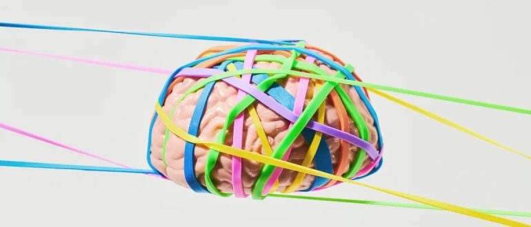
Brain wrapped in rubbberbands to demonstrate neuroplasticity

Does Brain Plasticity Increase After a Head Injury?

Medically reviewed by Nancy Hammond, M.D.
By James Roland for healthline.com on July 28, 2022

 
Brain plasticity, also called neuroplasticity, refers to the brain’s ability to adapt its structure and function in response to changes, such as a head injury or aging. Brain plasticity also involves the formation of new connections between neurons (brain cells).

The brain’s ability to reorganize these features after an injury affects the nature of post-injury recovery.

The severity of the injury goes a long way toward determining how the brain responds. But it’s often possible to boost brain plasticity with interventions and rehabilitation during the healing process.

What is brain plasticity?
Brain plasticity is a term that refers to the brain’s ability to restructure and reconfigure itself in response to change.

Change that can influence the brain comes in several forms. Expected changes include learning, experience, and aging. Unexpected changes include things like stroke and head injury.

Neuroplasticity has long been observed in children. It involves a process called neurogenesis, which is the formation of new neurons in the brain (and elsewhere in the nervous system).

There are two basic types of brain plasticity: structural and functional.

Structural Plasticity
Structural plasticity refers to the way the brain’s physical structure changes in response to learning.

For example, a small 2018 studyTrusted Source showed that healthy adults who participated in balance training twice a week, for 12 weeks, experienced thickening in certain areas of the brain involved in spatial orientation.

A 2016 study examined neuroplasticity in people learning to read Braille. It found that over the course of daily lessons, for 3 weeks, study participants developed increased connectivity in regions of the brain involved in processing sensations like touch.

Functional Plasticity
Functional plasticity refers to the brain’s ability to heal itself after injury. To achieve this, healthy regions of the brain adapt to take over certain functions that the damaged parts of the brain used to perform. This makes functional plasticity especially relevant for people recovering from head injuries.

A 2017 review of studies examining the role of neuroplasticity in stroke recovery found that a stroke can actually trigger neuroplasticity in certain areas. Neuroplasticity plays a role as the brain tries to resume regular functions, like speaking and controlling the movement of limbs.
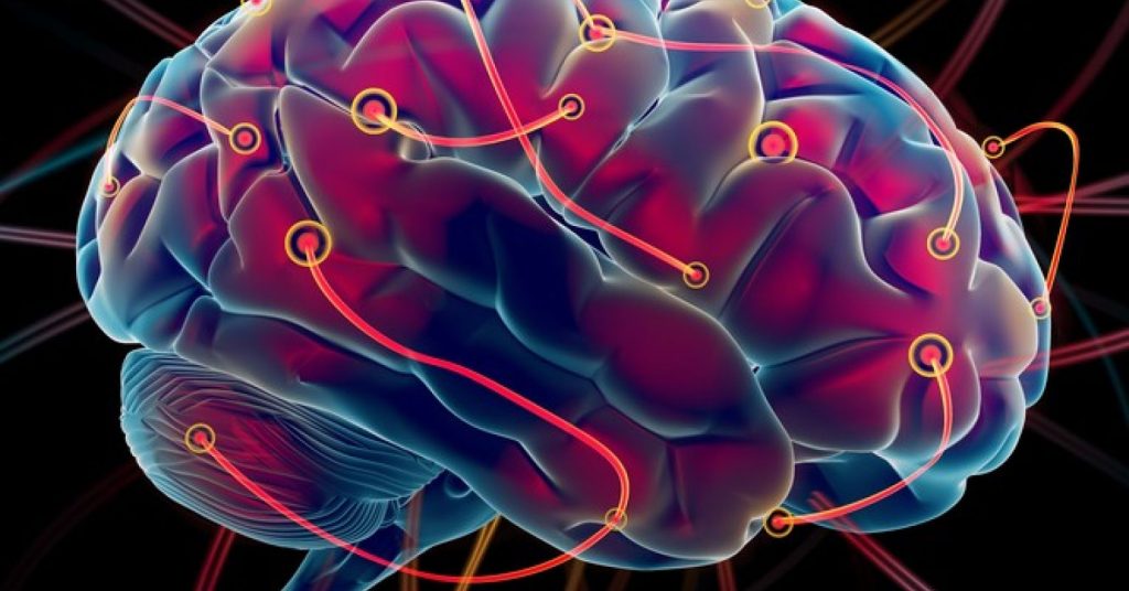
 
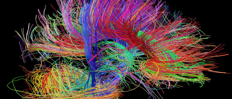
Brain image with new wiring

Can brain plasticity help you heal after a TBI?

 
A traumatic brain injury (TBI) refers to changes in brain function or brain health caused by an external force, such as a serious blow to the head.

The Centers for Disease Control and Prevention (CDC)Trusted Source reports that there were more than 220,000 TBI-related hospitalizations in 2019 and more than 64,000 TBI-related deaths the following year.

A TBI differs from a nontraumatic brain injury, also known as an acquired brain injury. Acquired brain injuries are those caused by internal factors, such as a stroke, which can damage brain tissue and affect muscle control, speech, cognition, and other functions.

When spontaneous brain plasticity doesn’t occur, it’s sometimes possible to boost neuroplasticity artificially.

 
A 2020 review of neuroplasticity therapies to treat stroke survivors suggests that approaches such as brain stimulation therapy and virtual reality might help enhance brain plasticity. It may also be possible to transfer nerves from healthy parts of the brain to injured parts.

Similarly, a 2017 review of studies on cognitive rehabilitation following TBI, suggests that memory and other thinking skills may be recovered to some degree with the help of cognitive rehabilitation. The studies showed how cognitive rehabilitation helped to modify damaged neural connections and various brain functions.

Does a brain injury increase neuroplasticity?

Because different regions of the brain are responsible for different functions, the location and severity of an injury determine which functions are affected and to what degree.

For example, certain areas of the brain are responsible for your ability to move certain parts of the body, like your left arm or your right foot.

This is where brain plasticity can help you heal after a brain injury. Just as exercise and learning can enhance brain structure and function, the body’s natural healing and recovery process after an injury can also increase neuroplasticity.

When neurons die due to injury, the brain naturally responds within a few days by developing new neural networks and recruiting various types of cells to take the place of those damaged or killed in the injury.

The extent to which neuroplasticity occurs depends on an individual’s age, the location of the injury, and other factors.

Does age matter after brain injury?

Whether it’s a brain injury or a broken wrist, being younger is always an advantage when it comes to recovery.

A 2008 studyTrusted Source of TBI survivors noted that disability scores following a TBI tended to be significantly better among younger TBI survivors compared with older individuals, even when those older survivors had less severe injuries. And the younger patients improved more in the first 5 years after the injury.

A 2019 report notes that because age affects neuroplasticity, the need for more strategies and therapies to compensate for age-related changes should be a higher priority in the face of an aging population.

Can you see brain plasticity on an MRI?

One of the most useful tools in diagnosing the impact of a TBI, stroke, or other injury or illness affecting the brain is magnetic resonance imaging (MRI).

An MRI can detect many changes in brain structure and function. Current technology is far from perfect, but it’s continuing to improve.

A 2021 articleTrusted Source suggests that advanced MRI techniques are helping doctors develop a more accurate picture of mild TBIs. This may help improve the treatment and understanding of mild TBIs in the future.

A newer type of MRI, called functional MRI (fMRI), can help doctors observe brain activity, not just brain structure. This may be particularly helpful in studying brain damage and recovery.

A 2017 studyTrusted Source of neuroimaging after TBI notes that fMRI can detect changes in thinking skills, emotions, and the course of neuroplasticity after an injury to the brain. The study says that fMRI is a helpful tool in assessing the damage caused by TBI and tracking brain changes during recovery.

But fMRI, the study says, will need to be accompanied by other data if it’s going to inform treatment decisions. This includes information gathered during cognitive-behavioral evaluations and other assessments.
Image Neurons reconnecting

How long does it take to heal after a TBI?

 
The time necessary to heal from a TBI can vary considerably from one person to the next. This is based mostly on the seriousness of the injury, as well as its location, the age of the individual, and that person’s overall physical and mental health.

A full recovery from a mild TBI can be expected in about 3 months. People with a moderate TBI will take longer to heal and will typically need cognitive rehabilitation, physical therapy, and other interventions.

Predicting the degree and length of recovery from a severe TBI is very difficult, and should be done on a case-by-case basis.

Takeaway

Brain plasticity after a head injury is when brain functions thought to be lost due to damage begin to be adopted by other, healthy brain tissue.

While not all functions can be reorganized or reestablished completely, the brain’s remarkable adaptability can often help people who had a stroke, traumatic brain injury, or other harmful events recover some function.

Brain plasticity can be encouraged through cognitive therapy, physical therapy, and other treatments.

CLICK HERE to read the original article
 

Brain image (Photo/Courtesy of USC Stevens Institute for Neuroimaging and Informatics)

Brain image (Photo/Courtesy of USC Stevens Institute for Neuroimaging and Informatics)

Researchers Create Maps of the Brain After Traumatic Brain Injury

Anne Warde, UC Irvine, June 17, 2022

 
Scientists from the University of California, Irvine have discovered that an injury to one part of the brain changes the connections between nerve cells across the entire brain.

The new research was published this week in Nature Communications.

Every year in the United States, nearly two million Americans sustain a traumatic brain injury (TBI). Survivors can live with lifelong physical, cognitive and emotional disabilities. Currently, there are no treatments.

One of the biggest challenges for neuroscientists has been to fully understand how a TBI alters the cross-talk between different cells and brain regions.

In the new study, researchers improved upon a process called iDISCO, which uses solvents to make biological samples transparent. The process leaves behind a fully intact brain that can be illuminated with lasers and imaged in 3D with specialized microscopes.

With the enhanced brain clearing processes, the UCI team mapped neural connections throughout the entire brain. The researchers focused on connections to inhibitory neurons, because these neurons are extremely vulnerable to dying after a brain injury. The team first looked at the hippocampus, a brain region responsible for learning and memory.

Then, they investigated the prefrontal cortex, a brain region that works together with hippocampus. In both cases, the imaging showed that inhibitory neurons gain many more connections from neighboring nerve cells after TBI, but they become disconnected from the rest of the brain.

“We’ve known for a long time that the communication between different brain cells can change very dramatically after an injury,” said Robert Hunt, PhD, associate professor of anatomy and neurobiology and director of the Epilepsy Research Center at UCI School of Medicine whose lab conducted the study, “But, we haven’t been able to see what happens in the whole brain until now.”

To get a closer look at the damaged brain connections, Hunt and his team devised a technique for reversing the clearing procedure and probing the brain with traditional anatomical approaches.

The findings surprisingly showed that the long projections of distant nerve cells were still present in the damaged brain, but they no longer formed connections with inhibitory neurons.

“It looks like the entire brain is being carefully rewired to accommodate for the damage, regardless of whether there was direct injury to the region or not,” explained Alexa Tierno, a graduate student and co-first author of the study. “But different parts of the brain probably aren’t working together quite as well as they did before the injury.”

The researchers then wanted to determine if it was possible for inhibitory neurons to be reconnected with distant brain regions.

To find out, Hunt and his team transplanted new interneurons into the damaged hippocampus and mapped their connections, based on the team’s earlier research demonstrating interneuron transplantation can improve memory and stop seizures in mice with TBI.

The new neurons received appropriate connections from all over the brain. While this may mean it could be possible to entice the injured brain to repair these lost connections on its own, Hunt said learning how transplanted interneurons integrate into damaged brain circuits is essential for any future attempt to use these cells for brain repair.

One of the biggest challenges for neuroscientists has been to fully understand how a TBI alters the cross-talk between different cells and brain regions. Image is in the public domain One of the biggest challenges for neuroscientists has been to fully understand how a TBI alters the cross-talk between different cells and brain regions. Image is in the public domain

“Our study is a very important addition to our understanding of how inhibitory progenitors can one day be used therapeutically for the treatment of TBI, epilepsy or other brain disorders,” said Hunt.

“Some people have proposed interneuron transplantation might rejuvenate the brain by releasing unknown substances to boost innate regenerative capacity, but we’re finding the new neurons are really being hard wired into the brain.”

Hunt hopes to eventually develop cell therapy for people with TBI and epilepsy. The UCI team is now repeating the experiments using inhibitory neurons produced from human stem cells.

“This work takes us one step closer to a future cell-based therapy for people,” Hunt said, “Understanding the kinds of plasticity that exists after an injury will help us rebuild the injured brain with a very high degree of precision. However, it is very important that we proceed step wise toward this goal, and that takes time.”

Jan C. Frankowski, PhD; Shreya Pavani; Quincy Cao and David C. Lyon, PhD also contributed to this study.

CLICK HERE to read the original article
 

Fatigue After Brain Injury

By Katherine Dumsa, OTR/L, CBIS and Angela Spears, MA, CCC-SLP, DPNS, CBIS, Rainbow Rehabilitation Centers

 
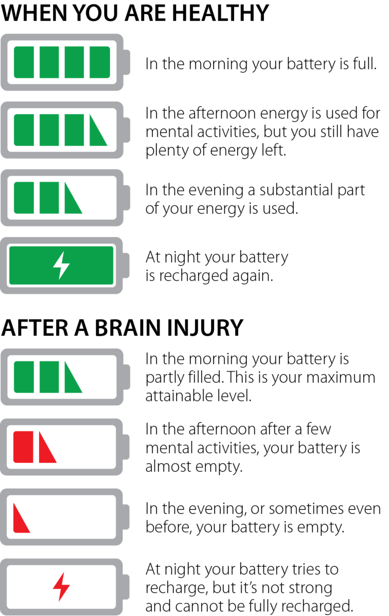
Fatigue is a part of life that is experienced by everyone. Whether it is from a busy day at work, a demanding workout, or after paying attention to a long lecture, the term “I’m tired” is exceedingly common.

Fatigue and Traumatic Brain Injuries
For individuals with brain injuries, fatigue (sometimes referred to as cognitive fatigue, mental fatigue, or neurofatigue), is one of the most common and debilitating symptoms experienced during the recovery process. It can become a significant barrier to one’s ability to participate in the activities they want and need to do in daily life. It is reported that as many as 98% of people who have experienced a traumatic brain injury have some form of fatigue. Many report that fatigue is their most challenging symptom after brain injury. Reasons for the fatigue are not well understood but may include endocrine abnormalities, the need for the brain to work harder to compensate for brain injury deficits (in other words, inefficiency), or changes to brain structures.

Assessment Tools to Determine Fatigue Levels
Fatigue can be difficult to identify because it is not always reported by the patient or obvious to others. Clinicians use various self-report assessment tools to gain further information on a patient’s fatigue levels and the impact it has on their overall daily functioning. Two of the scales specifically designed for individual patients with brain injuries include the Barrow Neurological Institute Fatigue Scale (BNI) and the Cause of Fatigue Questionnaire (COF). Clinicians must also evaluate physical and mental changes, which can lead to depression and other psychiatric conditions following brain injury. The changes can commonly present as overwhelming fatigue.

Symptoms
Generally, those who have sustained brain injuries have described fatigue as a sense of mental or physical tiredness, exhaustion, lack of energy, and/or low vitality. Physical observations of fatigue include yawning, an appearance of confusion or “brain fog,” or easily losing attention and concentration. In more severe cases, it may present as forgetfulness, irritability, slurred speech, or dizziness. Emotions can become raw at this level of fatigue, affecting mood, motivation, and interaction with one’s social network. To manage fatigue effectively, individuals must learn to identify the symptoms of fatigue and how to modify activities that may trigger fatigue. Managing fatigue effectively will help decrease stress levels and improve overall performance for both work and home activities. Some fatigue-inducing activities include:

  • Working at a computer
  • Watching television excessively
  • Having a stimulating sensory environment
  • Concentrating on paperwork
  • Reading for long periods of time
  • Physically demanding tasks
  • Cognitively demanding tasks
  • Emotionally draining tasks
  •  
    Symptoms of fatigue can include:

  • Physical Symptoms: a pale or greyish pallor, glazed eyes, headaches, tension in muscles, shortness of breath, slower movement and speech, decreased coordination, or difficulty staying awake.
  • Cognitive Symptoms: increased forgetfulness, distractibility, decreased ability to follow directions, making an increased number of mistakes, decreased awareness of surroundings, or increased response time or lack of response.
  • Social/Emotional Symptoms: decreased ability to communicate effectively, decreased ability to engage in social activities, irritability, restlessness, emotional lability, increased negative thoughts, withdrawal, short answers, dull tone of voice, lack of motivation and interest, or difficulty engaging in activities of daily living.
  •  
    Fatigue Is Not Laziness
    In today’s multi-media society, we take in, absorb, and process large amounts of information every day. It can be difficult for family members or peers to understand the limitations caused by fatigue following a brain injury. Unfortunately, it can be mistaken for laziness or an unwillingness to participate in therapies and daily activities. It is important to understand that lacking the mental energy needed to complete tasks does not equate to lacking the desire to complete those tasks. Many individuals struggling with fatigue have motivation but lack the energy to keep up with daily demands.

    Coping Strategies Used to Ease Symptoms
    When managing fatigue, it is important to identify and treat physical factors that may be contributing to the fatigue. Recognizing early signs of fatigue and working with the patient so they understand how to respond to these is beneficial. By learning to recognize these triggers, one can learn coping strategies to successfully meet daily demands, ultimately increasing quality of life. These strategies include:

  • Having a Healthy Sleep Routine – This can be done by setting a sleep schedule of when to go to bed and when to wake, regardless of the day of the week. Establishing a strict routine using an alarm clock allows the brain proper rest. When rest is needed, aim for a “power nap” of 30 minutes maximum to avoid feeling over tired for the remainder of the day. Lack of sleep has a negative effect on our cognition, mood, energy levels, and appetite. The American Academy of Neurology reports that as many as 40% to 65% of people with mild traumatic brain injury complain of insomnia, so maintaining a sleep hygiene program is essential to recovery and to managing fatigue.
  • Practicing Energy Conservation – Pacing yourself each day, or prioritizing daily tasks to avoid becoming over-tired, can help with balancing out a busy schedule. Complete tasks that require the most mental effort earlier in the day with planned rest breaks in the afternoon or evening.
    Organizing daily activities – Utilize a checklist or planner to set a to-do list. Break up complex projects into manageable tasks. When completing these tasks, minimize environmental stimulation as much as possible.
  • Improving Health and Wellness – Increased overall health and wellness has been described as “energizing,” and research suggests that it can improve mood. Aim to exercise three to five times per week for a minimum of 30 minutes per session. Maintain a well-balanced diet rich in protein, fiber, and carbohydrates to help the brain and body stay fully energized.
  • Keeping a Fatigue Diary – This kind of diary can assist in monitoring changes and energy levels before and after daily activities. This tracking of fatigue can be used with your treatment team to help mitigate what may be increasing neurofatigue. Assessment and treatment of fatigue continues to be a challenge for clinicians and researchers. While there is no cure for fatigue, there are many ways to manage and overcome the symptom. Awareness and an open mind towards coping strategies will lessen the negative effects of fatigue and allow for meaningful participation in life.
     
    REFERENCES

  • Keough, A. 2016. Strategies to manage neuro-fatigue.
  • Cantor, J.H., Ashman, T., Gordon, W., Ginsberg, A., Engmann, C., Egan, M., Spielman, L., Dijkers, M., & Flanagan, S. (2008). Fatigue after traumatic brain injury and its impact on participation and quality of life. Journal of Head Trauma Rehabilitation. 23(1), 41-51.
  • Jang, S., & Kwon, H. (2016). Injury of the ascending reticular activating system in patients with fatigue and hypersomnia following mild traumatic brain injury. Medicine. 95(6). e2628.
  • Belmont, A., Agar, N., Hugeron, B. Gallais, C. & Azouvi, P., Fatigue and traumatic brain injury. Annales de Réadaptation et de Médecine Physique. 49(6). 370-374.
  • Johnson, G. (2000). Traumatic brain injury survival guide. Traverse City, MI.
  • Heins, J., Sevat, R., Werkhoven, C. (n.d.) Neurofatigue. Brain Injury Explanation.
  •  
    This article first appeared in the Summer 2018 issue of Rainbow Visions Magazine available at www.rainbowrehab.com.

    CLICK HERE to read the original article
     

    Doctors Discuss Knowing the Signs of Concussion in Young Athletes

    By Adria Goins and Alex Onken, KSLA

     
    Thousands of students in the Arkansas/Louisiana/Texas (and across the nation) began fall sports over the last few weeks.

    However, with the new season here, comes a risk of injury. Football is the leading sport when it comes to concussions.

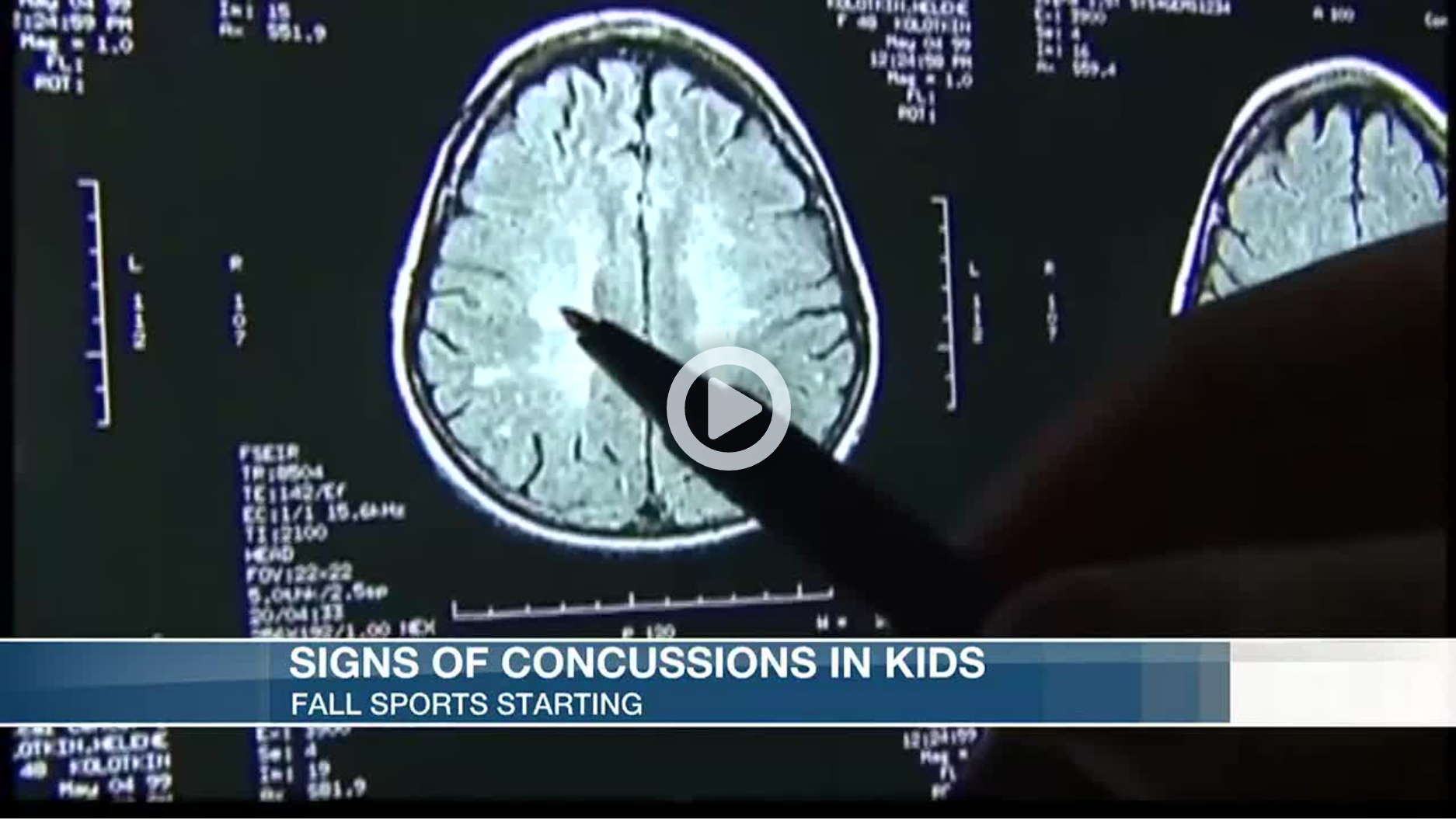
    The signs of a concussion are headache, fatigue and nausea. Parents are advised to then bring their child to a doctor right away if suspecting a possible concussion.

    “First diagnose it early and then after you diagnose it early make sure you avoid the triggers. So avoid extra screen time, over-exercising and just basically have 24 to 48 hours of cognitive physical rest,” said Dr. Kenneth Aguirre of Oschner-LSU Health Shreveport, who specializes in sports medicine.

    According to Dr. Charles Webb, also with Oschner-LSU Health and a sports medicine specialist, the topic of concussions and the potential risks of football comes up often.

    “I get that question a lot from parents. They want to know is it safe for my child to play high school or junior high, or even pee wee or popcorn football. and the question comes up because parents are worried about concussions. So my answer to them is if it were my child I wouldn’t let them play until they had an organized professional coach teaching them both how to hit and receive a hit.”

    Young athletes are usually taught how to hit and receive a hit around junior high. Dr. Webb said parents should put their children in club soccer or flag football in contrast to popcorn or pee wee football.

    “It’s much safer and you’re less likely to get hit in the head,” he said. “And you still get all the conditioning you need to play football later on in life.”

    In addition, doctors say keeping children awake when they have a concussion is a common misconception. Sleep is actually very good for the healing process.
     

    CLICK HERE to read the original article
     

    Best Practices for Managing Stress and Anxiety During Times of Uncertainty

    By Gary Seale, Ph.D., Regional Director, Centre for Neuro Skills

     
    The COVID-19 outbreak has produced a great deal of uncertainty and unwelcome anxiety. It’s no wonder we feel distressed when our daily routines have been severely disrupted. Due to social distancing and business closures, most people are not able to visit their favorite restaurant, go shopping, or engage in a work-out routine at the fitness center. When you couple this with concerns about personal health, uncertainty about how long these changes will last, and information and directives that change daily, it’s no wonder that many of us feel diseased or anxious. Several credible sites have recently posted simple but effective strategies to manage stress and remain emotionally healthy during this time of uncertainty. Some of the most frequent suggestions by experts at the American Psychological Association, Forbes, the Harvard Business Review, HealthLine, and others include the following:

    Differentiate between what is within your control vs. what is not in your control. Stay focused on the things you can do. Make a list and practice these regularly, and reward yourself for these practices. For example, you can:

  • Wash your hands, cover coughs and sneezes, etc.
  • Limit exposure to news. Manage stress by reducing (or eliminating) the number of times you check in to your favorite media outlet. The American Psychological Association recommends avoiding negative news right before bedtime.
  • Take care of your health (take your vitamins, get enough sleep, hydrate, engage in a daily exercise routine, etc.).
  • Practice your preferred relaxation technique if you feel stressed, such as deep breathing, yoga, mindful meditation, etc. If you don’t have a relaxation practice, now is a great time to develop one.
  •  

    Do the things that help you feel safe, such as:

  • Practice “social distancing” and limit exposure to groups of 10 or more people.
  • Give an “elbow bump” vs. a hug or a handshake.
  • Order out and have food delivered to your home or work; shop on-line and have items delivered to your home vs. going to a store or the mall.
  •  

    Rather than worrying about something that might happen in the future, stay focused on the present; maintain proper perspective.

  • Stay present and in the moment; focus on the task at hand.
  • If you feel yourself “borrowing trouble,” bring yourself back to the present.
  • Use a mindfulness practice (savor a favorite snack or meal; closely observe a pleasing object, such as a flower, etc.). If you don’t have a mindfulness practice, now is a great time to explore/develop one.
  • Put the situation into proper perspective (for example, as of March 24th in Houston/Harris County there are about 206 confirmed/presumed cases of COVID-19 and 2 deaths from this virus. Nineteen have fully recovered. Houston/Harris County has a population of approximately 6+ million. That translates into an infection statistic of 0.003%). Additionally, we live in a time and a nation of great abundance – an abundance of intellectual horsepower (some of the best minds in infectious diseases are here in the U.S. and are working on vaccines for this virus); an abundance of resources (the government has released over a trillion dollars to support research, product development and distribution, and aid to businesses); an abundance of entrepreneurs (companies re-tooling to overproduce cleaning supplies, ventilators, etc.).
  • This situation is temporary. As with other pandemics (MARS, SARS, H1N1, Zika, etc.), this too shall pass.
  •  

    Engage in proven positive psychology practices.

  • Start a gratitude list. Of all the positive emotions, gratitude is one of the most powerful and protective (from depressive symptoms). Simply start a list of all the things you are grateful for, large and small
  • Three Good Things. At the end of the day, list 3 good things that happened during the day. Once you have your list on paper, think about how you made those good things happen and reward yourself
  • Downward comparisons. Rather than thinking or saying to yourself, “I wish I…”, think or say to yourself, “I’m glad I…” For example, “I’m glad I work in the healthcare industry. I have a stable job, and in my work, I am able to help others.”
  • Connect with something larger than yourself. In the example above, you may find yourself feeling compassion for those out of work due to COVID-19, like servers in the restaurant industry. Ordering out from your favorite local restaurant and tipping big may help keep that business open during a time when the restaurant is closed to the public. Or you may want to visit a local blood bank and give blood as most all blood donation activities at large venues, like businesses, churches, etc. have been suspended for the time being.
  •  

    Get outside and enjoy nature.

  • During this time when we are practicing “social distancing”, we may feel “cooped up” which can result in feelings of anxiety. Getting outdoors and enjoying nature can lift your mood and help with managing stress. Walking increases heart rate and respiration, which is good for brain function and overall health. Spending time in the sunlight produces vitamin D, which improves the immune response. Being in “awe” (of nature) is another, very powerful positive emotion that can lift the mood and protect against depressive symptoms.
  •  

    Stay connected and don’t be afraid to ask for help.

  • Take this time to call friends and family and catch up.
  • Talk to a trusted friend about how you are feeling and all the practices you are using to stay healthy, both physically and mentally.
  • If you are feeling particularly distressed, reach out to a counselor or other mental health practitioner.
  • Talk to your supervisor or HR representative about your particular situation and any support you might need.
  •  

    Engage in resilience practices.

  • Think about a time when you faced a challenging situation and overcame it.
  • In your home or office, post some inspirational quotes, for example: “Never, never, never give up.” – Winston Churchill; “Failure is not an option.” – Gene Kranz, NASA Flight Director; “Tough times don’t last, but tough people do.” – Robert Schuller
  • Create a “resilience library” with inspirational books, videos, etc. For example, the book, “Unbroken” by Laura Hillenbrand, or the movie, “Remember the Titans”.
  • When possible, find “positives” (i.e., lower gas prices, safer commutes to work/less drunk drivers on the road, etc.), or lessons learned from the situation.
  • Use humor as appropriate.
  • If you have one, engage in your spiritual practice.
  •  

    CLICK HERE to read the original article
     

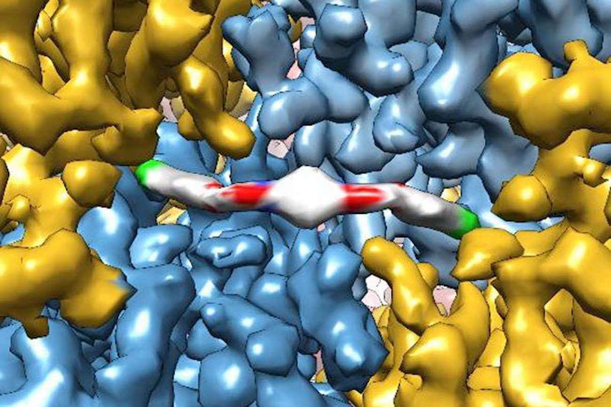
    ISRIB molecule—image by the Adam Frost lab at UCSF

    Drug Reverses Age-Related Mental Decline Within Days, Suggesting Lost Cognitive Ability is Not Permanent

    By Good News Network, December 27, 2020

     
    Just a few doses of an experimental drug that reboots protein production in cells can reverse age-related declines in memory and mental flexibility in mice, according to a new study by UC San Francisco scientists.

    The drug, called ISRIB, has already been shown in laboratory studies to restore memory function months after traumatic brain injury (TBI), reverse cognitive impairments in Down Syndrome, prevent noise-related hearing loss, fight certain types of prostate cancer, and even enhance cognition in healthy animals.

    In the new study, published Dec. 1 in the open-access journal eLife, researchers showed rapid restoration of youthful cognitive abilities in aged mice, accompanied by a rejuvenation of brain and immune cells that could help explain improvements in brain function—and with no side effects observed.

    “ISRIB’s extremely rapid effects show for the first time that a significant component of age-related cognitive losses may be caused by a kind of reversible physiological “blockage” rather than more permanent degradation,” said Susanna Rosi, PhD, Lewis and Ruth Cozen Chair II and professor in the departments of Neurological Surgery and of Physical Therapy and Rehabilitation Science.

    “The data suggest that the aged brain has not permanently lost essential cognitive capacities, as was commonly assumed, but rather that these cognitive resources are still there but have been somehow blocked, trapped by a vicious cycle of cellular stress,” added Peter Walter, PhD, a professor in the UCSF Department of Biochemistry and Biophysics and a Howard Hughes Medical Institute investigator. “Our work with ISRIB demonstrates a way to break that cycle and restore cognitive abilities that had become walled off over time.”

    Rebooting cellular protein production holds key to aging

    Walter has won numerous scientific awards, including the Breakthrough, Lasker and Shaw prizes, for his decades-long studies of cellular stress responses. ISRIB, discovered in 2013 in Walter’s lab, works by rebooting cells’ protein production machinery after it gets throttled by one of these stress responses—a cellular quality control mechanism called the integrated stress response (ISR; ISRIB stands for ISR InhiBitor).

    The ISR normally detects problems with protein production in a cell—a potential sign of viral infection or cancer-promoting gene mutations—and responds by putting the brakes on cell’s protein-synthesis machinery. This safety mechanism is critical for weeding out misbehaving cells, but if stuck in the ‘on’ position in a tissue like the brain, it can lead to serious problems, as cells lose the ability to perform their normal activities, according to Walter and colleagues.

    In particular, their recent animal studies have implicated chronic ISR activation in the persistent cognitive and behavioral deficits seen in patients after TBI, by showing that, in mice, brief ISRIB treatment can reboot the ISR and restore normal brain function almost overnight.

    The cognitive deficits in TBI patients are often likened to premature aging, which led Rosi and Walter to wonder if the ISR could also underlie purely age-related cognitive decline. Aging is well known to compromise cellular protein production across the body, as life’s many insults pile up and stressors like chronic inflammation wear away at cells, potentially leading to widespread activation of the ISR.

    “We’ve seen how ISRIB restores cognition in animals with traumatic brain injury, which in many ways is like a sped-up version of age-related cognitive decline,” said Rosi, who is director of neurocognitive research in the UCSF Brain and Spinal Injury Center and a member of the UCSF Weill Institute for Neurosciences. “It may seem like a crazy idea, but asking whether the drug could reverse symptoms of aging itself was just a logical next step.”

    Signature effects of aging disappeared literally overnight

    In the new study, researchers led by Rosi lab postdoc Karen Krukowski, PhD, trained aged animals to escape from a watery maze by finding a hidden platform, a task that is typically hard for older animals to learn. But animals who received small daily doses of ISRIB during the three-day training process were able to accomplish the task as well as youthful mice—and much better than animals of the same age who didn’t receive the drug.

    The researchers then tested how long this cognitive rejuvenation lasted and whether it could generalize to other cognitive skills. Several weeks after the initial ISRIB treatment, they trained the same mice to find their way out of a maze whose exit changed daily—a test of mental flexibility for aged mice who, like humans, tend to get increasingly stuck in their ways. The mice who had received brief ISRIB treatment three weeks before still performed at youthful levels, while untreated mice continued to struggle.

    To understand how ISRIB might be improving brain function, the researchers studied the activity and anatomy of cells in the hippocampus, a brain region with a key role in learning and memory, just one day after giving animals a single dose of ISRIB. They found that common signatures of neuronal aging disappeared literally overnight: neurons’ electrical activity became more sprightly and responsive to stimulation, and cells showed more robust connectivity with cells around them while also showing an ability to form stable connections with one another usually only seen in younger mice.

    The researchers are continuing to study exactly how the ISR disrupts cognition in aging and other conditions and to understand how long ISRIB’s cognitive benefits may last. Among other puzzles raised by the new findings is the discovery that ISRIB also alters the function of the immune system’s T cells, which also are prone to age-related dysfunction. The findings suggest another path by which the drug could be improving cognition in aged animals, and could have implications for diseases from Alzheimer’s to diabetes that have been linked to heightened inflammation caused by an aging immune system.

    “This was very exciting to me because we know that aging has a profound and persistent effect on T cells and that these changes can affect brain function in the hippocampus,” said Rosi. “At the moment, this is just an interesting observation, but it gives us a very exciting set of biological puzzles to solve.”

    Success shows the ‘serendipity’ of basic research

    Rosi and Walter were introduced by neuroscientist Regis Kelly, PhD, executive director of the University of California’s QB3 biotech innovation hub, following Walter’s 2013 study showing that the drug seemed to instantly enhance cognitive abilities in healthy mice. To Rosi, the results from that study implied some walled-off cognitive potential in the brain that the molecule was somehow unlocking, and she wondered if this extra cognitive boost might benefit patients with neurological damage from traumatic brain injury.

    The labs joined forces to study the question in mice, and were astounded by what they found. ISRIB didn’t just make up for some of the cognitive deficits in mice with traumatic brain injury—it erased them. “This had never been seen before,” Rosi said. “The mantra in the field was that brain damage is permanent—irreversible. How could a single treatment with a small molecule make them disappear overnight?”

    Further studies demonstrated that neurons throughout the brains of animals with traumatic brain injury are thoroughly jammed up by the ISR. Using ISRIB to release those brakes lets brain cells immediately get back to their normal business. More recently, studies in animals with very mild repetitive brain injury—akin to pro athletes who experience many mild concussions over many years—showed that ISRIB could reverse increased risk-taking behavior associated with damage to self-control circuits in the frontal cortex.

    “It’s not often that you find a drug candidate that shows so much potential and promise,” Walter says, calling it “just amazing”.

    No side effects

    One might think that interfering with the ISR, a critical cellular safety mechanism, would be sure to have serious side effects, but so far in all their studies, the researchers have observed none. This is likely due to two factors. First, it takes just a few doses of ISRIB to reset unhealthy, chronic ISR activation back to a healthier state. Second, ISRIB has virtually no effect when applied to cells actively employing the ISR in its most powerful form—against an aggressive viral infection, for example.

    ISRIB has been licensed by Calico, a South San Francisco, Calif. company exploring the biology of aging, and the idea of targeting the ISR to treat disease has been picked up by many other pharmaceutical companies, Walter says.

    “It almost seems too good to be true, but with ISRIB we seem to have hit a sweet spot for manipulating the ISR with an ideal therapeutic window,” Walter said.

    Get more links to background studies from original article from UCSF News.
     

    CLICK HERE to read the original article


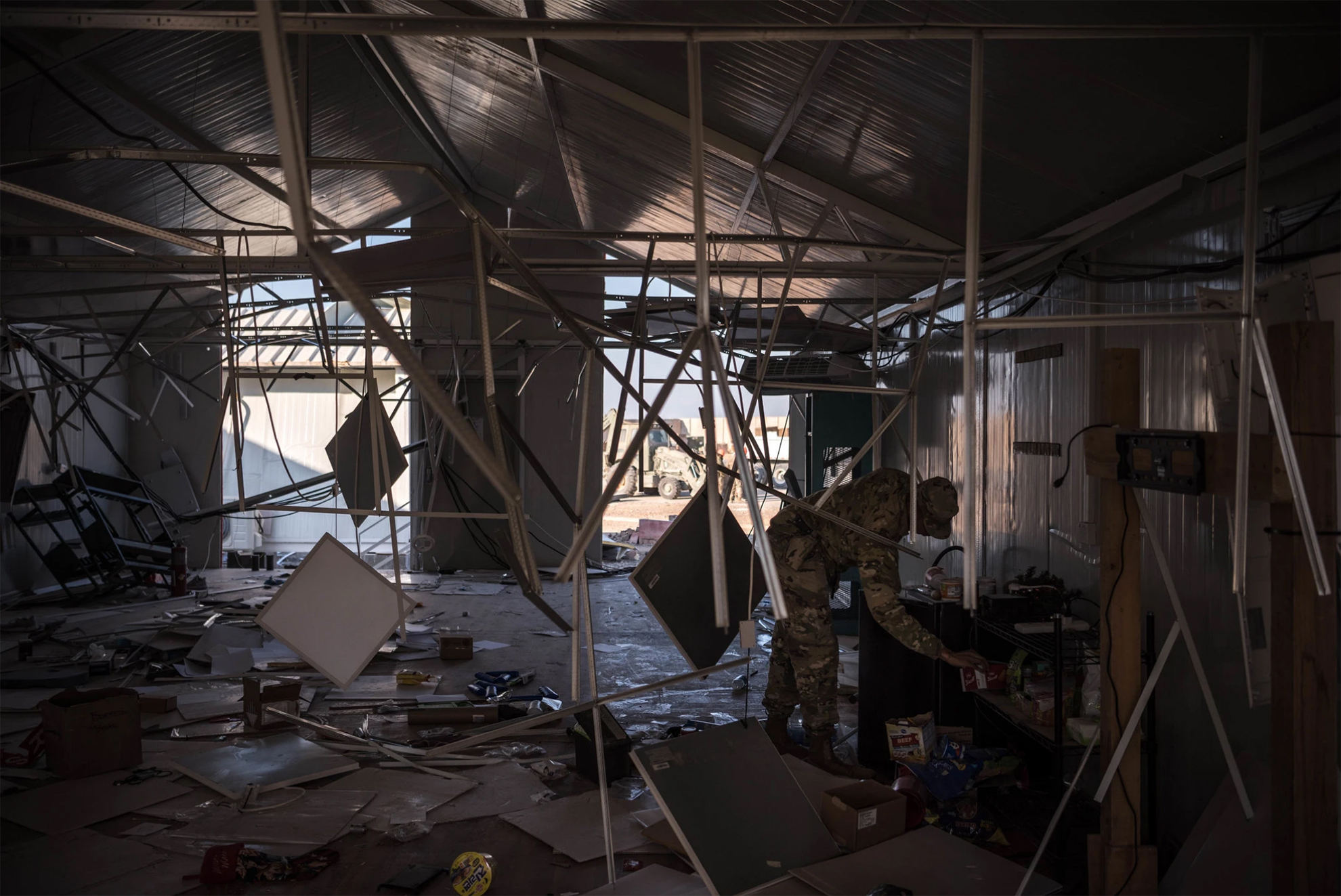
    Ayn al Asad Air Base in western Iraq after an Iranian missile attack on Jan. 8. The number of service members experiencing symptoms associated with brain injuries has since topped 100. Photo Credit…Sergey Ponomarev for The New York Times


    Brain Injuries Are Common in Battle.
    The Military Has No Reliable Test for Them.

    Traumatic brain injury is a signature wound of the wars in Iraq and Afghanistan. But the military still has no objective way of diagnosing it in the field.

    By Dave Philipps and Thomas Gibbons-Neff for nytimes.com, February 15, 2020

     
    U.S. troops at Ayn al Asad Air Base in western Iraq hunkered down in concrete bunkers last month as Iranian missile strikes rocked the runway, destroying guard towers, hangars and buildings used to fly drones.
    When the dust settled, President Trump and military officials declared that no one had been killed or wounded during the attack. That would soon change.

    A week after the blast, Defense Department officials acknowledged that 11 service members had tested positive for traumatic brain injury, or TBI, and had been evacuated to Kuwait and Germany for more screening. Two weeks after the blast, the Pentagon announced that 34 service members were experiencing symptoms associated with brain injuries, and that an additional seven had been evacuated. By the end of January the number of potential brain injuries had climbed to 50. This week it grew to 109.

    The Defense Department says the numbers are driven by an abundance of caution. It noted that 70 percent of those who tested positive for a TBI had since returned to duty. But experts in the brain injury field said the delayed response and confusion were primarily caused by a problem both the military and civilian world have struggled with for more than a decade: There is no reliable way to determine who has a brain injury and who does not.

    Top military leaders have for years called traumatic brain injury one of the signature wounds of the wars in Iraq and Afghanistan; at the height of the Iraq war in 2008, they started pouring hundreds of millions of dollars into research on detection and treatment. But the military still has no objective tool for diagnosing brain injury in the field. Instead, medical personnel continue to use a paper questionnaire that relies on answers from patients — patients who may have reasons to hide or exaggerate symptoms, or who may be too shaken to answer questions accurately.

    The military has long struggled with how to address so-called invisible war wounds, including traumatic brain injury and post-traumatic stress disorder. Despite big investments in research that have yielded advances in the laboratory, troops on the ground are still being assessed with the same blunt tools that have been in use for generations.

    The problem is not unique to the military. Civilian doctors struggle to accurately assess brain injuries, and still rely on a process that grades the severity of a head injury in part by asking patients a series of questions: Did they black out? Do they have memory problems or dizziness? Are they experiencing irritability or difficulty concentrating?

    “It’s bad, bad, bad. You would never diagnose a heart attack or even a broken bone that way,” said Dr. Jeff Bazarian a professor of emergency medicine at the University of Rochester Medical Center. “And yet we are doing it for an injury to the most complex organ in the body. Here’s how crazy it gets: You are relying on people to report what happened. But the part of the brain most often affected by a traumatic brain injury is memory. We get a lot of false positives and false negatives.”

    Without a good diagnosis, he said, doctors often don’t know whether a patient has a minor concussion that might require a day’s rest, or a life-threatening brain bleed, let alone potential long-term effects like depression and personality disorder.

    At Ayn al Asad, personnel used the same paper questionnaires that field medics used in remote infantry platoons in 2010. Aaron Hepps, who was a Navy corpsman in a Marines infantry company in Afghanistan at that time, said it did not work well then for lesser cases, and the injuries of many Marines may have been missed. During and after his deployment, he counted brain injuries in roughly 350 Marines — about a third of the battalion.

    After the January missile attack, Maj. Robert Hales, one of the top medical providers at the air base, said that the initial tests were “a good start,” but that it took numerous screenings and awareness among the troops to realize that repeated exposure to blast waves during the hourlong missile strikes had affected dozens.

    Traumatic brain injuries are among the most common injuries of the wars in Iraq and Afghanistan, in part because armor to protect from bullet and shrapnel wounds has gotten better, but they offer little protection from the shock waves of explosions. More than 350,000 brain injuries have been reported in the military since 2001.

    The concrete bunkers scattered around bases like Ain al Assad protect from flying shrapnel and debris, but the small quarters can amplify shock waves and lead to head trauma.

    The blasts on Jan. 8, one military official said, were hundreds of times more powerful than the rocket and mortar attacks regularly aimed at U.S. bases, causing at least one concrete wall to collapse atop a bunker with people inside.

    Capt. Geoff Hansen was in a Humvee at Ayn al Asad when the first missile hit, blowing open a door. Then a second missile hit.

    “That kind of blew me back in,” he said. “Blew debris in my face so I went and sat back down a little confused.”

    A tangle of factors make diagnosing head injuries in the military particularly tricky, experts say. Some troops try to hide symptoms so they can stay on duty, or avoid being perceived as weak. Others may play up or even invent symptoms that can make them eligible for the Purple Heart medal or valuable veteran’s education and medical benefits.

    And sometimes commanders suspect troops with legitimate injuries of malingering and force them to return to duty. Pentagon officials said privately this week that some of the injuries from the Jan. 8 incident had probably been exaggerated. Mr. Trump seemed to dismiss the injuries at a news conference in Davos, Switzerland, last month. “I heard they had headaches,” he said. “I don’t consider them very serious injuries relative to other injuries I have seen.”

    In the early years of the war in Iraq, troops with concussions were often given little medical treatment and were not eligible for the Purple Heart. It was only after clearly wounded troops began complaining of poor treatment that Congress got involved and military leaders began pressing for better diagnostic technology.

    Damir Janigro, who directed cerebrovascular research at the Cleveland Clinic for more than a decade, said relying on the questionnaire makes accurate diagnosing extremely difficult.

    “You have the problem of the cheaters, and the problem of the ones who don’t want to be counted,” he said. “But you have a third problem, which is that even if people are being completely honest, you still don’t know who is really injured.”

    In civilian emergency rooms, the uncertainty leads doctors to approve unnecessary CT scans, which can detect bleeding and other damage to the brain, but are expensive and expose patients to radiation. At the same time doctors miss other patients who may need care. In a war zone, bad calls can endanger lives, as troops are either needlessly airlifted or kept in the field when they cannot think straight.

    Mr. Janigro is at work on a possible solution. He and his team have developed a test that uses proteins found in a patient’s saliva to diagnose brain injuries. Other groups are developing a blood test.

    Both tests work on a similar principle. When the brain is hit by a blast wave or a blow to the head, brain cells are stretched and damaged. Those cells then dispose of the damaged parts, which are composed of distinctive proteins. Abnormal levels of those proteins are dumped into the bloodstream, where for several hours they can be detected in both the blood and saliva. Both tests, and another test being developed that measures electrical activity in the brain, were funded in part by federal grants, and have shown strong results in clinical trials. Researchers say they could be approved for use by the F.D.A. in the next few years.

    The saliva test being developed by Mr. Janigro will look a bit like an over-the-counter pregnancy test. Patients with suspected brain injuries would put sensors in their mouths, and within minutes get a message that says that their brain protein levels are normal, or that they should see a doctor.

    But the new generation of testing tools may fall short, said Dr. Gerald Grant, a professor of neurosurgery at Stanford University and a former Air Force lieutenant colonel who frequently treated head injuries while deployed to Iraq in 2005.

    Even sophisticated devices had trouble picking up injuries from roadside bombs, he said.

    “You’d get kids coming in with blast injuries,” he said, “and they clearly had symptoms, but the CT scans would be negative.”

    He was part of an earlier effort to find a definitive blood test, which he said in an interview was “the holy grail.” But progress was slow. The grail was never found, he said, and the tests currently being developed are helpful for triaging cases, but too vague to be revolutionary.

    “Battlefield injuries are complex,” he said. “We still haven’t found the magic biomarker.”

    CLICK HERE to go to the original article


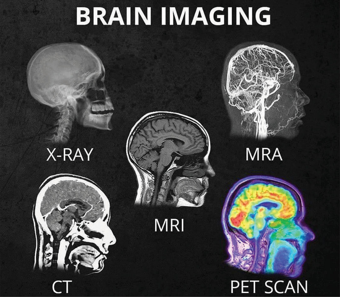
    What’s the difference between all the different head scans (X-Ray, CT, MRI, MRA, PET scan)? And what do they show in the head?

    Michael S. Tehrani, M.D.Follow Founder & CEO at MedWell Medical

     
    Ever wonder what’s the difference between all the different head scans (xray, CT, MRI, MRA, PET scan) and what they show in the head. Well wonder no more. The Dr. T easy to understand version…

    X-Ray: shows bone/skull only. Does not show the brain. Best used to detect if there are bone fractures.

    CT: a quick test. Shows brain but detail not great. Shows if any larger bleed, stroke, lesions, or masses.

    MRI: a long test. Shows brain and detail is great. Shows smaller bleeds, stroke, lesions, or masses.

    MRA:
    shows the flow of blood in the vasculature system of the brain. If there is vessel narrowing or blockage this test would show it.

    PET scan: shows how active different parts of the brain is. An active brain uses sugar as energy and pet scan detects how much sugar is being used by lighting up and turning different colors. The more sugar being used the more that area will light up and be different in colors. Cancer cells use the most sugar so cancer cells light up the most. PET scan is used to see if there are cancer cells. (Cancer cells replicate at a very fast and uncontrolled rate hence use a lot of sugar to allow that replication hence why they light up so much).

    CLICK HERE to download the original article


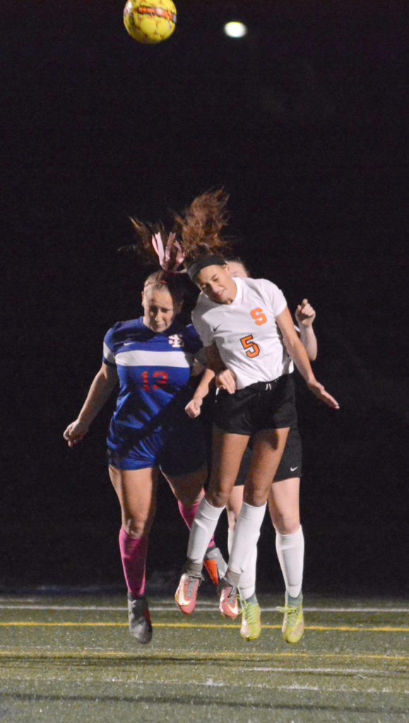
    High school injury reports analyzed by InvestigateWest and Pamplin Media show that girls are twice as likely to get concussions as boys in Oregon. Girls in the 13U age group, pictured above, are the youngest allowed to use headers.
     

    The Concussion Gap: Head injuries in girls soccer are an ‘Unpublicized Epidemic’

    Lee van der Voo, InvestigateWest, photos by David Ball / Pamplin Media Group

     
    When it comes to concussion in sports, all eyes are on football, or so it seems. But it’s not just football that causes a high number of head injuries among young athletes.

    Another culprit? Girls soccer.

    National research has found girls are more likely to suffer a concussion than boys in any sport. In 2017, researchers at Northwestern University generated national headlines when they found concussion rates among young female soccer players were nearly as high as concussion rates for boys playing football — and roughly triple the rate of concussions in boys soccer.

    In Oregon, injury reports from public high schools analyzed by InvestigateWest and Pamplin Media Group mirrored that trend, showing soccer concussions were second to those from football between 2015 and 2017. What’s more, at the schools that included the gender of injured athletes, there were nearly twice as many reports of possible concussions for girls playing soccer than boys in the sport.

    The rate of concussions in girls soccer worries local experts like Jim Chesnutt, a doctor in sports medicine at Oregon Health & Science University, who says those injuries are not widely recognized, even as concussion rates rise for girls playing soccer.

    “In a lot of ways, it’s a growing epidemic for young girls that I think has gone unpublicized,” said Chesnutt, co-director of the Oregon Concussion Awareness and Management Program and a member of the Governor’s Task Force on Traumatic Brain Injury.

    More exposure, more injury

    It’s understandable that much of the youth concussion conversation centers on football, given the physical contact that is visibly — and audibly — evident on every play, as well as the large rosters and the lengthy lists of players who are injured.

    But if you compare girls soccer with football, and only look at the high school participation and injury data, “you’re missing a gigantic part of the picture,” according to Michael Koester, a doctor of sports medicine at the Slocum Center in Eugene. He directs its sports concussion program and serves as the chair of the Sports Medicine Advisory Committee for the National Federation of State High School Associations.

    Koester notes that high school boys play eight to 10 football games per season, and typically play other sports in the off-season.

    Girls, however, play 15 to 20 soccer games in a high school season, but when that season ends, they may play another 80-plus games throughout the winter, spring and summer with club teams, said Koester, who, like Chesnutt, is a medical adviser to the Oregon Schools Activities Association.

    “If we’re looking at injury risk by athletic exposure,” which is one practice or game, a standard in evaluating risk, Koester said, female soccer players probably are playing five if not 10 times more practices and games than football players.

    And Koester doesn’t see the trend ending.

    “The thought used to be that this was all revolving around, ‘Wow! They want to get their kid a scholarship,’ ” he said. “Now it’s kind of gotten to the point where there’s so much single-sport participation that we see kids that are specializing in sport early, just so they’ll be able to make their high school team.”

    Single-sport athletes are more prone to injury in any sport. According to a study by scientists at the University of Wisconsin, high school athletes who specialized in just one sport at an early age were twice as likely to suffer injuries to their lower extremities.

    “We see a lot of overuse injury among girls playing soccer,” Koester said. “We see a lot of ACL injury among girls playing soccer. It’s a well-known problem.”

    Aggressive play

    Another factor is the evolution of sports.

    Angella Bond is an athletic trainer for Tuality Sports Medicine and works on the sidelines with athletes at Hillsboro schools. Anecdotally, she said, all athletes push to be bigger, faster and stronger. Soccer is no exception, nor are girls.

    As athletes develop, they take bigger hits at higher speeds, and competitive games build on their momentum. As competition grows in girls soccer, the sport is trending to be more aggressive, she said.

    “Unfortunately, I think that happens with girls sports,” she said. “Arms fly a little bit more.”

    Chesnutt agreed. “I think over the years, soccer has become more physical,” he said. “And I think the physical contact and the aggressive nature of that physical contact is more associated with concussions.”

    According to the American Academy of Pediatrics, soccer — unlike football, ice hockey and lacrosse — is not a “collision sport.” But it is a “contact sport” because athletes “routinely make contact with each other or inanimate objects.”

    Header balls, though often singled out as a source of concussions, are not necessarily to blame.

    The force created when a soccer ball meets a head can rattle a brain, but data increasingly points to other factors when competitors vie for a ball in the air.

    According to a study by The Research Institute at Nationwide Children’s Hospital, while headers accounted for 27 percent of concussions, it was knocks with other players on aerial play — including head-to-head contact and arms and elbows to the head — and contact with the ground that accounted for 70 percent of those concussions in girls soccer, suggesting aggressive play is a factor in most concussions involving headers.

    Why girls?

    But why are girls more prone to concussions than boys while playing soccer? The prevailing theories focus on their weaker neck-muscle development, weaker body strength (needed to stabilize the neck and head during aerial play), and more frequent contact with the ground. A year ago, a study in the Journal of the American Osteopathic Association found that female high school soccer players took twice as long as male players to recover.

    It’s also possible that girls don’t benefit as much from early treatment. A recent study published by the American Academy of Pediatrics found that girls are five times more likely than boys to stay on the pitch and play through a head injury.

    And the soccer community has been slow to recognize the hard hits its girls are taking. Instead, soccer is at the forefront of the cultural empowerment of girls.

    Local experts concerned about concussion risk note that sports, including girls soccer, have plenty of benefits. Just being physically active is good for kids, and sports like soccer help establish lifelong fitness habits, teach team-building skills, and promote character development and assertiveness.

    “The worry is that the take-home message is that (girls soccer) is healthy and fantastic and nothing can be bad about it,” said Koester, who says an opposite negative message, equally extreme, is more often associated with boys playing football.

    Greater awareness needed

    Concussion education and awareness in girls soccer is paramount, according to local experts such as Chesnutt.

    “I think the way to decrease it is to really analyze how we can modify the amount of body contact that goes on in soccer to limit the dangerous aggressive behavior that is associated with concussion,” he said.

    Unlike youth football, a sport that’s adjusting to new information about concussions all the time, soccer has largely failed to address new information about concussions, Chesnutt said.

    Football, for example, has reduced head-to-head helmet play, limited full-contact practices and games, and zeroed in on the specialty teams with the highest concussion rates.

    “Football has really done, I think, an exceptional job of identifying some areas where there have been some definite higher incidents and some problems,” said Chesnutt, who lectures nationally about youth concussions. “As a group of coaches, leagues, parents and referees, they’ve all looked at it and come up with some solutions that have decreased concussion rates. And I think it’s time for soccer to do the same thing.”

    Read the original article
     

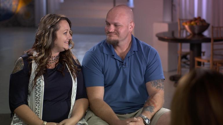
    Libby and Tom Bates // CBS News

    A brain disease best known for impacting football players who suffered concussions is now being found in soldiers

    By Sharyn Alfonsi, September 16, 2018, CBS News

    Until a few years ago, NFL players who struggled with severe depression, bouts of rage and memory loss in their retirement were often told they were just having a hard time adjusting to life away from the game. Doctors have since learned these changes can be symptoms of the degenerative brain disease CTE – chronic traumatic encephalopathy, caused by blows to the head.

    As we first reported in January, CTE isn’t just affecting athletes, but also showing up in our nation’s heroes. Since 9/11 over 300,000 soldiers have returned home with brain injuries. Researchers fear the impact of CTE could cripple a generation of warriors.

    When Joy Kieffer buried her 34-year old son this past summer, it was the end of a long goodbye.

    Kieffer’s son, Sgt. Kevin Ash, enlisted in the Army Reserves at the age of 18. Over three deployments, he was exposed to 12 combat blasts, many of them roadside bombs. He returned home in 2012 a different man.

    Joy Kieffer: His whole personality had changed. I thought it was exposure to all of the things that he had seen, and he had just become harder. You know, but he was — he was not happy.

    Sharyn Alfonsi: So at this point, you’re thinking this decline, this change in my child is just that he’s been in war and he’s seen too much.

    Joy Kieffer: Right.

    Sharyn Alfonsi: Did he tell you about blasts that he experienced during that time?

    Joy Kieffer: Uh-huh.

    Sharyn Alfonsi: What did he–tell you?

    Joy Kieffer: That they shook him. And he was having blackouts. And — it frightened him.

    Ash withdrew from family and friends. He was angry. Depressed. Doctors prescribed therapy and medication, but his health began to decline quickly. By his 34th birthday, Sgt. Kevin Ash was unable to speak, walk or eat on his own.

    Sharyn Alfonsi: Looking back on it now, was there anything you feel like he could’ve done?

    Joy Kieffer: Uh-uh.

    Sharyn Alfonsi: Because?

    Joy Kieffer: Because it was– it– it was his brain. The thing I didn’t know was that his brain was continuing to die. I mean, before he went into the service he said, “you know, I could come back with no legs, or no arms, or even blind, or I could be shot, I could die,” but nobody ever said that he could lose his mind one day at a time.

    His final wish was to serve his country one last time by donating his brain to science — a gesture he thought would bring better understanding to the invisible wounds of war.

    Joy reached out to the VA-Boston University-Concussion Legacy Foundation Brain Bank where neuropathologist Dr. Ann McKee is leading the charge in researching head trauma and the degenerative brain disease CTE.

    McKee has spent fourteen years looking at the postmortem brains of hundreds of athletes who suffered concussions while playing their sport.

    Last summer, her findings shook the football world when she discovered CTE in the brains of 110 out of 111 deceased NFL players — raising serious concerns for those in the game today.

    And when Dr. McKee autopsied Patriots tight-end Aaron Hernandez who killed himself after being convicted of murder, she found the most severe case of CTE ever, in someone under 30.

    Now she’s seeing similar patterns in deceased veterans who experienced a different kind of head trauma — combat blasts. Of the 125 veterans’ brains Dr. Mckee’s examined, 74 had CTE.

    Sharyn Alfonsi: I can understand a football player who keeps, you know, hitting his head, and having impact and concussions. But how is it that a combat veteran, who maybe just experienced a blast, has the same type of injury?

    Dr. Ann McKee: This blast injury causes a tremendous sort of– ricochet or– or– a whiplash injury to the brain inside the skull and that’s what gives rise to the same changes that we see in football players, as in military veterans.

    Blast trauma was first recognized back in World War I. Known as ‘shell shock,’ poorly protected soldiers often died immediately or went on to suffer physical and psychological symptoms. Today, sophisticated armor allows more soldiers to walk away from an explosion but exposure can still damage the brain — an injury that can worsen over time.

    Dr. Ann McKee: It’s not a new injury. But what’s been really stumping us, I think, as– as physicians is it’s not easily detectable, right? It’s– you’ve got a lot of psychiatric symptoms– and you can’t see it very well on images of the brain and so it didn’t occur to us. And I think that’s been the gap, really, that this has been what everyone calls an invisible injury.

    Dr. Ann McKee: This is the world’s largest CTE brain bank.

    The only foolproof way to diagnose CTE is by testing a post-mortem brain.

    Sharyn Alfonsi: So these are full of hundreds of brains…

    Dr. Ann McKee: Hundreds of brains, thousands really…

    Researchers carefully dissect sections of the brain where they look for changes in the folds of the frontal lobes – an area responsible for memory, judgement, emotions, impulse control and personality.

    Dr. Ann McKee: Do you see there’s a tiny little hole there? That is an abnormality. And it’s a clear abnormality.

    Sharyn Alfonsi: And what would that affect?

    Dr. Ann McKee: Well, it’s part of the memory circuit. You can see that clear hole there that shouldn’t be there. It’s connecting the important memory regions of the brain with other regions. So that is a sign of CTE.

    Thin slivers of the affected areas are then stained and viewed microscopically. It’s in these final stages where a diagnosis becomes clear as in the case of Sgt. Kevin Ash.

    Sharyn Alfonsi: So this is Sergeant Ash’s brain?

    Dr. Ann McKee: Right. This is– four sections of his brain. And what you can see is– these lesions. The, and those lesions are CTE And they’re in very characteristic parts of the brain. They’re at the bottom of the crevice. That’s a unique feature of CTE.

    Sharyn Alfonsi: And in a healthy brain, you wouldn’t see any of those kind of brown spots?

    Dr. Ann McKee: No, no, it would be completely clear. And then when you look microscopically, you can see that the tau, which is staining brown and is inside nerve cells is surrounding these little vessels.

    Sharyn Alfonsi: And explain, what is the tau?

    Dr. Ann McKee: So tau is a protein that’s normally in the nerve cell. It helps with structure and after trauma, it starts clumping up as a toxin inside the nerve cell. And over time, and even years, gradually that nerve cell dies.

    Dr. Lee Goldstein has been building on Dr. McKee’s work with testing on mice.

    Inside his Boston University lab, Dr. Goldstein built a 27-foot blast tube where a mouse – and in this demonstration, a model – is exposed to an explosion equivalent to the IEDs used in Iraq and Afghanistan.

    Dr. Lee Goldstein: When it reaches about 25 this thing is going to go.

    Dr. Goldstein’s model shows what’s going on inside the brain during a blast. The brightly colored waves illustrate stress on the soft tissues of the brain as it ricochets back and forth within the skull.

    Dr. Lee Goldstein: What we see after these blast exposures, the animals actually look fine. Which is shocking to us. So they come out of what is a near lethal blast exposure, just like our military service men and women do. And they appear to be fine. But what we know is that that brain is not the same after that exposure as it was microseconds before. And if there is a subsequent exposure, that change will be accelerated. And ultimately, this triggers a neurodegenerative disease. And, in fact, we can see that really after even one of these exposures.

    Sharyn Alfonsi: The Department of Defense estimates hundreds of thousands of soldiers have experienced a blast like this. What does that tell you?

    Dr. Lee Goldstein: This is a disease and a problem that we’re going to be dealing with for decades. And it’s a huge public health problem. It’s a huge problem for the Veterans Administration. It’s a huge moral responsibility for all of us.

    A responsibility owed to soldiers like 34-year-old Sgt. Tom Bates.

    Sgt. Tom Bates: We were struck with a large IED. It was a total devastation strike.

    Bates miraculously walked away from a mangled humvee — one of four IED blasts he survived during deployments in Iraq and Afghanistan.

    Sharyn Alfonsi: Do you remember feeling the impact in your body?

    Sgt. Tom Bates: Yes. Yeah.

    Sharyn Alfonsi: What does that feel like?

    Sgt. Tom Bates: Just basically like getting hit by a train.

    Sharyn Alfonsi: And you were put back on the frontlines.

    Sgt. Tom Bates: Yes.

    Sharyn Alfonsi: And that was it?

    Sgt. Tom Bates: Uh-huh

    When Bates returned home in 2009, his wife Libby immediately saw a dramatic change.

    Libby Bates: I thought, “Something is not absolutely right here. Something’s going on. For him to just lay there and to sob and be so sad. You know, what do you do for that? How do I– how do I help him? He would look at me and say, “If it wasn’t for you, I would end it all right now.” You know, I mean, like, what do you– what do you do– and what do you say to somebody who says that? You know I love this man so much. And —

    Sharyn Alfonsi: You’re going to the VA, you’re getting help, but did you feel like you weren’t getting answers?

    Sgt. Tom Bates: Yes.

    Sharyn Alfonsi: And so you took it into your own hands and started researching?

    Sgt. Tom Bates: I knew the way everything had gone and how quick a lot of my neurological issues had progressed that something was wrong. And I just– I wanted answers for it.

    That led him to New York’s Mount Sinai Hospital where neurologist Dr. Sam Gandy is trying to move beyond diagnosing CTE only in the dead by using scans that test for the disease in the living.

    Dr. Sam Gandy: By having this during life, this now gives us for the first time the possibility of estimating the true prevalence of the disease. It’s important to estimate prevalence so that people can have some sense of what the risk is.

    In the past year, 50 veterans and athletes have been tested for the disease here. Tom Bates asked to be a part of it.

    That radioactive tracer – known as t807 – clings to those dead clusters of protein known as tau, which are typical markers of the disease.

    Through the course of a 20 minute PET scan, high resolution images are taken of the brain and then combined with MRI results to get a 360 degree picture of whether there are potential signs of CTE.

    Scan results confirmed what Tom and Libby had long suspected.

    On the right, we see a normal brain scan with no signs of CTE next to Tom’s brain where tau deposits, possible markers of CTE, are bright orange.

    Dr. Sam Gandy: Here these could be responsible for some of the anxiety and depression he’s suffered and we’re concerned it will progress.

    Sgt. Tom Bates: My hope is that this study becomes more prominent, and gets to more veterans, and stuff like that so we can actually get, like, a reflection of what population might actually have this.

    There is no cure for CTE.

    Dr. Gandy hopes his trial will lead to drug therapies so he can offer some relief to patients like Tom.

    Dr. Ann McKee believes some people may be at higher risk of getting the disease than others.

    While examining NFL star Aaron Hernandez’s brain she identified a genetic bio-marker she believes may have predisposed him to CTE.

    A discovery that could have far-reaching implications on the football field and battlefield.

    Sharyn Alfonsi: Do you think you will ever be your old self again?

    Sgt. Tom Bates: I don’t ever see me being my old self again. I think it’s just too far gone.

    Sharyn Alfonsi: So what’s your hope then?

    Sgt. Tom Bates: Just to not become worse than I am now.

    Since our story first aired, over 100 veterans have contacted Dr. Gandy to enroll in ongoing trials to identify whether they are living with CTE. And more than 300 have reached out to Dr. Mckee about donating their brains to research.

    Read the original article
     

    Serving the Brain Injury Community Since 1983