News

Junior Seau, shown at his beloved Pacific Ocean in the ESPN Films “30 for 30” documentary “Seau,” which premieres Thursday. (ESPN Films)

ESPN hits the mark with documentary ‘Seau’

By Tom Krasovic, September 20, 2018, San Diego Union Tribune

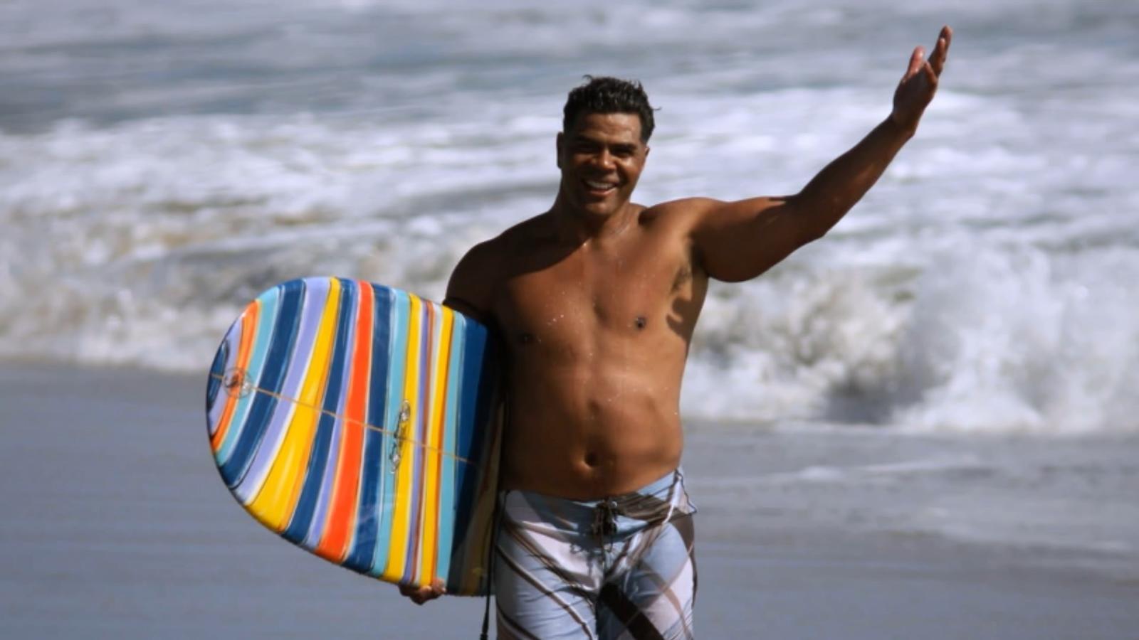
An aerial view of the Oceanside coast, in full sparkle and splendor below, grandly eases viewers into “Seau,” an ESPN Films documentary in the “30 for 30” series that debuts Thursday on the streaming service ESPN+.

It’s a sunny scene, the Pacific Ocean’s turquoise waves illuminated as they roll toward the white beach. The late Junior Seau told friends he found peace paddling on these waters, deep into his life alongside the town where he’d grown up.

Up at dawn with a yellow long-board and oar in hand, Seau had only a short walk from his beachfront home to the water.

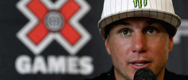
Yet the former Chargers linebacker, role model and local philanthropist was then also writing in a journal of bouts with depression, memory loss and perceived guilt. There were headaches, too, and nights plagued by insomnia. “Buddy,” he’d told a friend and professional soccer player who’d suffered a brain injury from heading a ball, “I’ve had a concussion since I was 15.”

Diary entries also revealed feelings of humiliation and embarrassment over not living up to expectations of others and himself, and of feeling used by others.

“The world has nothing for me,” Seau pens in one entry, the cursive words all too legible.

One of Seau’s surviving adult children, after reading the grim line aloud, wonders why his father didn’t regard his family as something in this apparent world of nothing.

Why couldn’t they have been a lifeline for him to reach out and grasp?

“Seau,” produced and directed by Kirby Bradley, lets viewers draw their own conclusions about a complicated life that ended one May morning six years ago, at age 43, with a self-inflicted gunshot wound to the chest, but not before we hear from an array of family members, friends and experts in football and brain science.

At the end of the 90-minute film, themes of redemption and hope are raised.

“Let’s all walk from here being better for having known Junior Seau and the impact he had on our lives,” NFL quarterback Drew Brees, a former Chargers teammate of the Hall of Fame linebacker, concludes near the film’s end.

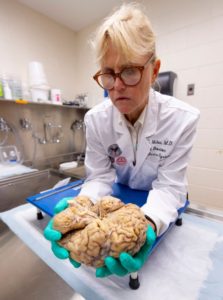
Former Chargers lineman Aaron Taylor notes that in death, Seau drew extraordinary attention to the link between head trauma and a degenerative brain disease, CTE, revealed in a tissue sample sent to a brain scientists at the family’s request.

Exciting beginnings and success are a thread to the film, followed often by bitter detours or hurtful endings.

Seau took to sports at Oceanside High with a passion that rivaled his stunning blend of size, speed and agility. If he was slamming into football ball-carriers or catching passes, scoring baskets or throwing the discus and shot, he was a “force of nature” for the green-and-white-clad Pirates, observers said.

A flood of football scholarship offers came to the small home where Seau and his brothers slept in a tiny garage.

Jubilation ensued when Seau chose USC, keeping him close to his parents and siblings and the tight-knit Samoan-American community in Oceanside. A similar celebration arose in 1990 when the Chargers drafted him fifth overall. “I’m a real momma’s boy,” Seau said, pulling on a blue team cap.

Playing for his beloved “Diego,” he led the long-struggling Chargers to the playoffs in just his third season, and their first Super Bowl two years later. “Now the world is gonna know the San Diego Chargers,” he told some 70,000 celebrants in Mission Valley after the team returned from claiming the 1994 AFC title in Pittsburgh.

The flip side?

If Oceanside lost a game in which he played, Junior lost his lunch money. It was the price his father exacted.

The thrill of signing with USC gave way to humiliation when a failed admittance test made him ineligible as a freshman. His father refused to talk to him in response, deeming the failure an embarrassment to the family. After a dominant junior year with USC, there would be no senior year. Making money was the next step, in no small part because he wanted to support his parents and other family members.

The Chargers couldn’t build upon their Super Bowl season, and the team’s constant losing wore on Seau.

When the Chargers traded him in the spring of 2003, after 13 seasons with the club, Seau was hurt that the team — Stay Unclassy, San Diego? — called not him but his agent to tell him the news. “I know that was hard on him,” said the agent, Steve Feldman.

Gina Seau was working for the Chargers in marketing when she first met Seau early in his NFL career.

She recalled “very kind eyes” and a “very soft voice” that almost “didn’t match the size and stature.”

The two would marry, but erratic behavior that Gina Seau linked to numerous football-related head injuries — “My head is on fire,” he told her — led to a divorce in 2002. The two remained friends. Believing that driving off a steep coastal cliff in October 2010 wasn’t an accident, Gina pleaded with her former husband to get help.

Here’s hoping that if there’s a “Seau II,” events yet to transpire bring more developments of redemption. Say, a cure for CTE.

Read the original article


Ann C. McKee, chief of neuropathology at the VA Boston Healthcare System, which houses the world’s largest brain bank devoted to CTE research, examines a brain earlier this month.(Photo: Robert Deutsch, USA TODAY)

Researchers close in on CTE diagnosis in living, one brain at a time

By Nancy Armour, August 24, 2018, USA TODAY

BOSTON – Submerged in chemicals in the stainless-steel bowl is the key to life and, researchers hope, death.

It’s a human brain. That of a man who played college football in the 1950s, to be exact. His family donated his brain to get answers for themselves, but what’s found could lead to more answers about chronic traumatic encephalopathy, the devastating neurodegenerative disease linked to concussions and repetitive head trauma from football and other contact sports.

“Our main objective, our overarching goal, is to help the people who are living. To be able to diagnose this disease during life,” says Ann McKee, chief of neuropathology at the VA Boston Healthcare System, which houses the world’s largest brain bank devoted to CTE research.

“If we can diagnose it, we can monitor it and test therapies to see if they’re effective in treating this disease,” says McKee, director of the CTE Center at Boston University’s School of Medicine. “It would really dramatically increase our ability to point out genetic susceptibilities for this. We’d be able to look at how much is too much in certain individuals or certain positions in certain sports.”

As another football season begins, it inevitably leads to questions and fears about head trauma and its long-term damage. How many hits are too many? What can parents do to protect their children or players do to protect themselves? Are athletes in certain sports more susceptible?

Most important, which athletes will develop CTE – or Parkinson’s or ALS (amyotrophic lateral sclerosis) – and why?

The answers will come from brains such as the one McKee dissected this month, when USA TODAY Sports toured the brain bank.

The brain bank has more than 500 brains, most of them donated by former athletes or their families who suspected CTE because of mood swings, behavioral changes, depression or dementia. Of those brains, more than 360 had CTE, McKee says.

SEARCHING FOR CLUES

The arrival of a brain sets two teams in motion. One set of clinicians talks to the family to find out more about the donors. Did they play any sports? If so, what and for how long? When did they start? Did they experience any other kind of head trauma, say from an automobile accident, domestic violence or military service? Did they have drug or alcohol problems? How did their mental health change, and when did that occur?

Separately, and usually without any information about the person whose brain it was, McKee and her researchers study the brain. It is cut in half, and one half is stored in a minus-80-degree freezer, so it will be available for molecular, genetic and biochemical studies.

The other half is then photographed and sectioned. After removing the brain stem, McKee uses what looks like a bread knife to cut slices of the brain about a quarter-inch thick.


Ann C. McKee slices the brain into segments about a quarter-inch thick as part of in-depth, time consuming research on the organ. McKee hopes the work will unlock answers to CTE. (Photo: Robert Deutsch, USA TODAY)
 
Simply by looking at the brain, McKee can tell a few things. The brain of this man, who was in his 80s when he died, has shrunk, noticeably smaller than it should be for a man who once played football. The folds of the brain, normally pressed tightly against one another, are loose and have gaps between them, some large enough that the tip of a finger could be inserted.

She points to the ventricles, chambers in the middle of his brain that are filled with fluid during life. They should be small, but these are “just gigantic.”

“As the brain shrinks, they expand. What this indicates is there’s been enormous shrinkage of the brain,” McKee says. “Those are huge.”

The hippocampus, a section in the middle of the brain that controls memory, is small but not abnormally so for a man in his 80s. If it was, that could be an indication of Alzheimer’s. But a membrane that runs from one side of the brain to the other, normally thick like a rubber band, has shrunk. In some spots, it’s almost invisible.

“This is looking more like frontal predominant atrophy, and that could mean CTE because Alzheimer’s almost always affects the hippocampus,” McKee says. “At this point, I always want to know, ‘What is it? Let’s look under the microscope.’ But you have to wait.”

CTE can’t be seen by the naked eye, and it takes at least three weeks to prepare slides of the brain tissue.


CTE is caused by tau, a protein in the brain released as a result of head trauma. When tau clumps together, it damages brain cells and can change the brain’s function. Though tau causes Alzheimer’s, McKee says, the tau that causes CTE looks distinctly different.

Under a microscope, it can be seen in telltale brown spots.

“CTE is very focal. In fact, in its early stages, it’s in the crevices. It just piles up. And that’s around blood vessels,” McKee says. “That’s very different. Alzheimer’s never does that.”

As CTE progresses, those clusters or clumps of tau will spread, and the disease will become more severe. That’s why, in the early stages of disease, stages 1 and 2, the symptoms usually relate to behavioral changes or mood swings. In stages 3 and 4, the disease is exhibited in memory loss.

“We think there may be more pathology in the young players than we’re appreciating just with the tau protein,” McKee says. “We think there’s maybe white matter structural changes or maybe inflammatory changes that are responsible for that loss of control, which is so difficult for the individuals.”

‘EVERY CASE IS A MYSTERY’

Once the slides have been examined, the pathologists and clinicians will come together for a conference. At this point, neither knows what the other does. The clinicians detail what they’ve learned about the brain donor’s history and suggest a diagnosis. The pathologists will then say whether the brain tissue confirms it.

“Every case is a mystery,” McKee says. “It’s not the same way you usually solve a mystery. I solve the pathology first, and then you go back and find out (the history). And then you try and put the two together.”

Some former players and their families once were reluctant to donate their brains, but that stigma largely has disappeared. So much so that McKee said brains arrive at the Boston bank almost every day.

Though that lengthens the time it takes to reach a definitive diagnosis, it will shorten the time before a living diagnosis can be found. In addition to the work done in her lab, McKee shares tissue samples with researchers around the world.

“What we want to do is establish the risk, educate people, educate parents, educate players,” McKee says. “So if they’re unwilling to risk that future self, if they’re unwilling to take that risk because it’s too high for them personally, we want to give them enough data so they can make a very sound and wise decision.”

When that day comes, it will change sports forever.

Read the original article
 

With better devices, science can get closer to a more complete picture of how neurons interact for cognitive functionality. (Photo/iStock)

Are we getting closer to a complete brain mapping? New devices explore more regions safely

By Breanne Grady, April 13, 2018, viterbischool.usc.edu

Researchers have developed thin, flexible polymer-based materials that record activity in more subregions of the brain with safer, more specific placement.

Science has yet to unravel a complete understanding of the brain and all its intricate workings. It’s not for lack of effort.

Over many decades, multiple research studies have sought to understand the dizzying “talk,” or interconnectivity, between thousands of microscopic entities in the brain, in particular neurons. The goal: to one day arrive at a complete brain “mapping” — a feat that could unlock tremendous therapeutic potential.

Researchers at the USC Viterbi School of Engineering have developed thin, flexible polymer-based materials for use in microelectrode arrays that record activity more deeply in the brain and with more specific placement than ever before. What’s more is that each microelectrode array is made up of eight “tines,” each with eight microelectrodes which can record from a total 64 subregions of the brain at once.

Same Quality, More Safety

In addition, the polymer-based material, called Parylene C, is less invasive and damaging to surrounding cells and tissue than previous devices comprised of silicon or microwires. However, the long and thin probes can easily buckle upon insertion, making it necessary to add a dissolvable brace made up of polyethylene glycol (PEG) that prevents it from bending.

Professor Ellis Meng of the USC Viterbi Department of Biomedical Engineering said that the performance of the new polymer-based material is on par with microwires in terms of recording fidelity and sensitivity. “The information that we can get out is equivalent, but the damage is much less,” Meng said. “Polymers are gentler on the brain, and because of that, these devices get recordings of neuronal communication over long periods of time.”

As with any prosthetic implant, caution must be exercised in terms of the body’s natural immune response to a foreign element. In addition to inflammation, previous microelectrode brain implants made of silicon or microwires have caused neuronal death and glial scarring, which is damage to connective tissue in the nervous system. However, Parylene C is biocompatible and can be microfabricated in extremely thin form to mold well to specific subregions of the brain, allowing for exploration with minimal damage.

Listening In

So far, these arrays have been used to record synaptic responses of individual neurons within the hippocampus, a part of the brain responsible for memory formation. If injured, the hippocampus may be compromised, resulting in a patient’s inability to form new memories. Meng, a faculty member of the Michelson Center for Convergent Bioscience, said that the polymer-based material can conform to a specific location in the hippocampus and “listen in on a conversation” between neurons. Because there are many such “eavesdroppers” (the microelectrodes), much more information about their interconnectivity can be gleaned.

“I can pick where I want my electrodes to be, so I can match up to the anatomy of the brain,” Meng, the Dwight C. and Hildagarde E. Baum Chair, said. “Along the length of a tine, I can put a group of electrodes here and a group of electrodes there, so if we plant to a certain depth, it’s going to be near the neurons I want to record from.”

Up Next

Future research will determine the recording lifetime of polymer-based arrays and their long-term “signal-to-noise” (SNR) stability. Also, the team plans to create devices with even higher density, including a double-sided microelectrode array with 64 electrodes per tine instead of eight — making for a total of around 4,000 electrodes placed in the brain at once.

In addition to Meng, Professor Ted Berger, the David Packard Chair in Engineering, and Research Professor Dong Song (both of the USC Viterbi School of Engineering) were co-authors along with Ph.D. students Huijing Xu and Ahuva Weltman Hirschberg and post-doctoral scholar Kee Scholten. Funding was provided be the National Science Foundation (NSF) and the National Institutes of Health (NIH). The study titled “Acute in vivo testing of a conformal polymer microelectrode array for multi-region hippocampal recordings” now published in the Journal of Neural Engineering.

About the Michelson Center

The USC Michelson Center for Convergent Bioscience brings together a diverse network of premier scientists and engineers under one roof, thanks to a generous $50 million gift from orthopedic spinal surgeon, inventor and philanthropist Gary K. Michelson, and his wife, Alya Michelson. At the Michelson Center, scientists and engineers from the USC Dornsife College of Letters, Arts and Sciences, USC Viterbi School of Engineering and Keck School of Medicine of USC are working to solve some of the greatest intractable problems of the 21st century in biomedical science, including a fundamentally new understanding of the cell and new approaches for cancer, neurological and cardiovascular disease.

Read the original article
 

The Intrepid Spirit traumatic brain injury treatment center is slated to open April 2 at Camp Pendleton. (Courtesy Naval Hospital Camp Pendleton) (Photo/iStock)

Brain injury center to open at Marine base

By Linda McIntosh, March 27, 2018, sandiegouniontribune.com

A brain injury treatment center for military personnel will open its doors April 2 near the Naval Hospital Camp Pendleton.

The $11.5 million Intrepid Spirit center is the seventh of nine such facilities at military bases across the country. It is funded by the New York-based nonprofit Intrepid Fallen Heroes Fund founded in 2000 by Zachary Fisher, who also started the Fisher House Foundation for military families.

The center will operate as a part of Naval Hospital Camp Pendleton to treat active-duty military patients who suffer from the physical and psychological effects of brain injury. The center will also provide education and other resources on brain injury for veterans and the wider community.

The center will expand the hospital’s existing program at the Concussion Care Clinic, which has served more than 2,000 patients since 2014. An estimated 550-600 new patients are expected to be referred to the center each year.

“The facility will offer interdisciplinary, state-of-the-art evaluation of service members using clinical, laboratory and imaging resources to guide treatment,” said Cmdr. Paul Sargent, medical director of the Intrepid Spirit center, Naval Hospital Camp Pendleton.

The center’s specialty rehabilitation and therapy programs will focus on providing service members strategies to improve recovery from physical, emotional and spiritual injuries.

“We know that being able to be close to home, surrounded by loved ones, is a crucial part of the recovery process, so we are opening centers on the West Coast this spring at Camp Pendleton and also at Joint Base Lewis-McChord in Washington in order that service members who need treatment do not have to uproot themselves and their families to get it,” said David Winters, president of the Intrepid Fallen Heroes Fund.

Two teams of clinicians will serve the clinic. Their specialties range from neurology, physical medicine and rehabilitation, psychiatry, trauma psychology, neuropsychology and pain psychology to physical and occupational therapy, creative arts therapy and neuro-optometry.

“Our approach is a broadly collaborative center for preventing, treating and researching head trauma and injury to the brain,” Sargent said.

The Intrepid Spirit center includes research, education and clinical staff from the Defense and Veterans Brain Injury Center, which is part of the Department of Defense’s Health Agency.

“Teaching Marines, sailors and their commands about the risks of head injury, how to mitigate concussions and how to understand Traumatic Brain Injury signs and symptoms, along with how to improve readiness is a major goal of our TBI training,” said Regional Education Coordinator Clint Pearman, a certified brain injury specialist with the Defense and Veterans Brain Injury Center.

Pearman provides outreach, education, training and resources for medical personnel, military commands, service members, veterans and family members and civilian community groups from the Camp Pendleton area up to northern California.

The center’s design is based on the original National Intrepid Center of Excellence, which opened in 2010 at the Walter Reed National Military Medical Center in Bethesda, Md., operated by the Department of Defense.

“There are hundreds of thousands of U.S. service members who continue to suffer from traumatic brian injury and other psychological health conditions,” Winters said. “The Intrepid Fallen Heroes Fund has tried to help these brave men and women get the best care available, so we made it our mission to build nine Intrepid Spirit centers that provide comprehensive, state-of-the-art treatment.”

The clinic’s ground breaking was last May and a grand opening ceremony will be held at 11 a.m. April 4 at the Intrepid Spirit Center.

For information about base access, visit pendleton.marines.mil/About/Base-Information/Base-Access.

Read the original article


The radial-arm water maze is a common test to assess working memory in rodents.

Memory-enhancing drug reverses effects of traumatic brain injury in mice

By Ryan Cross, Jul. 10, 2017, sciencemag.org

Whether caused by a car accident that slams your head into the dashboard or repeated blows to your cranium from high-contact sports, traumatic brain injury can be permanent. There are no drugs to reverse the cognitive decline and memory loss, and any surgical interventions must be carried out within hours to be effective, according to the current medical wisdom. But a compound previously used to enhance memory in mice may offer hope: Rodents who took it up to a month after a concussion had memory capabilities similar to those that had never been injured.

The study “offers a glimmer of hope for our traumatic brain injury patients,” says Cesario Borlongan, a neuroscientist who studies brain aging and repair at the University of South Florida in Tampa. Borlongan, who reviewed the new paper, notes that its findings are especially important in the clinic, where most rehabilitation focuses on improving motor—not cognitive—function.

Traumatic brain injuries, which cause cell death and inflammation in the brain, affect 2 million Americans each year. But the condition is difficult to study, in part because every fall, concussion, or blow to the head is different. Some result in bleeding and swelling, which must be treated immediately by drilling into the skull to relieve pressure. But under the microscope, even less severe cases appear to trigger an “integrated stress response,” which throws protein synthesis in neurons out of whack and may make long-term memory formation difficult.

In 2013, the lab of Peter Walter, a biochemist at the University of California, San Francisco (UCSF), discovered a compound—called ISRIB—that blocked the stress response in human cells in a dish. Surprisingly, when tested in healthy mice, ISRIB boosted their memory. Wondering whether the drug could also reverse memory impairment, Walter teamed up with UCSF neuroscientist Susanna Rosi to study mouse models of traumatic brain injury. First, they showed that the stress response remains active in the hippocampus, a brain region important for learning and memory, for at least 28 days in injured mice. And they wondered whether administering ISRIB would help.

Rosi and her team first used mechanical pistons to hit anesthetized mice in precise parts of their surgically exposed brains, resulting in contusive injuries, focused blows that can also result from car accidents or being hit with a heavy object. After 4 weeks of rest, Rosi trained the mice to swim through a water maze, where they used cues to remember the location of a hidden resting platform. Healthy mice got better with practice, but the injured ones didn’t improve. However, when the injured mice were given ISRIB 3 days in a row, they were able to solve the maze just as quickly as healthy mice up to a week later, the researchers report today in the Proceedings of the National Academy of Sciences.

“We kept replicating experiments, thinking maybe something went wrong,” Rosi says. So the team decided to study ISRIB in a second model of traumatic brain injury known as a closed head injury, which resembles a concussion from a fall. They again used a mechanical piston, but this time landed a broad blow to the back of the skull. Two weeks later, the mice were trained on a tougher maze, full of bright lights and loud noise. They had to scurry around a tabletop with 40 holes, looking for the one with an escape hatch. Again, while the uninjured mice improved at the task, the concussed mice never got the hang of it. But after four daily doses of ISRIB, the concussed mice performed as well as their healthy counterparts. “This is the most exciting piece of work I’ve ever done, no doubt,” Rosi says.

“Paradigm shift is not too strong a term to use,” says Ramon Diaz-Arrastia, neurologist and director of clinical traumatic brain injury research at the University of Pennsylvania. “This … shows for the first time that a therapy in the chronic period of traumatic brain injury can have pretty potent effects.” Walter agrees. “Normally you would give up on these mice and say nothing can be done here,” he says. “But ISRIB just magically brings the cognitive ability back.”

Still, Borlongan cautions that studies in animals often don’t pan out when tested in humans. He says that this drug has a leg up, though, because it was tested in two models and also readily crosses the blood-brain barrier, which prevents many drugs that look good on paper from entering the brain and having an effect.

If the therapy translates to humans, it could be a boon for soldiers returning from war, who sometimes wait weeks between leaving the battlefield and arriving home for treatment. Brian Head, a neurobiologist at the VA San Diego Healthcare System in California notes that traumatic brain injury is still hard to diagnose, especially with veterans that show up to the clinic long after the injury. “But right now nothing else is working, and giving a compound [that works] a month later is really impressive.”

In 2015, ISRIB was licensed to the secretive Google spinout company Calico, which studies the biology of aging and life span. Walter says his lab has a research agreement with Calico to pursue “basic mechanistic work” on ISRIB, but that the new study was not funded by Calico. Google declined to comment on the new research.

Although the protein target of ISRIB is known, the exact manner in which the drug restores memory is hazy. The team hypothesizes that ISRIB may work by allowing normal protein synthesis—essential for making new neuronal connections and thus forming new memories—to resume, which would otherwise be blunted by the integrated stress response. “Even if this drug doesn’t materialize, other ways of manipulating the integrated stress response may lead to an effective treatment in the future,” Walter says.

Read the original article
 

CARSON, CA – AUGUST 03: Bronze medal winner Dave Mirra speaks in a press conference after the Rally Car race during the summer X Games 14 at Home Depot Center on August 3, 2008 in Carson, California. (Photo by Christian Petersen/Getty Images)

Months after committing suicide, Dave Mirra has become the first action sports athlete to be diagnosed with CTE

by Robert Silverman, vocativ.com (May 24, 2016)
 
After BMX biking legend Dave Mirra committed suicide on February 4 of this year, his wife had his brain tested for chronic traumatic encephalopathy. Sadly, the result came back positive, rife with tau proteins dotting both his temporal and frontal lobes after years of enduring an unknown amount of concussive and sub-concussive trauma. This makes Mirra the first action sports athlete to be diagnosed with CTE.

The neuropathologist went so far as to equate the condition of his brain to that of NFL players and other contact sport athletes that have been posthumously diagnosed with the disease. “I couldn’t tell the difference,” Dr. Lili-Naz Hazrati said.

In an exclusive interview with ESPN: The Magazine, Mirra’s wife Lauren describes the agonizing final weeks of his life, the transformation of his formerly vibrant personality into something different and darker, prone to wild mood swings and unprovoked crying jags or bouts of exhaustion, his mind clouded and wracked with depression.

“I remember seeing him sitting on our bed one day, in the last month of his life,” she said. “I had just gotten out of the shower and saw him hunched over with the blankest lost look. I sat down next to him and held his hand. I said, ‘What is wrong? Are you OK?’ And he just shrugged his shoulders. He couldn’t even speak. He didn’t know. He couldn’t put it into words. He was lost. He was helpless. It was completely different from who he was.”

“He was gone. I could see straight through him,” she continued. “It was the hardest thing to see, looking at someone you love, and you can’t have a conversation with them, and you can see straight through their eyes.”

Lauren Mirra doesn’t know what her exact plans might be for the future, but her overarching hope is that she’ll be able to find a forum in which to speak out, to encourage best practices and prevention measures, without coming across as an ideologue out to ban action sports altogether.

“Through him we have an opportunity to help and change,” she said. “Beauty from ashes. That’s how I will always choose to see it.”

Read the original article


The Cohen family partners with USC to serve families in Los Angeles.
 
by Lynn Lipinski, tfm.USC.edu (Autumn 2016) — PEACE AFTER WAR can be elusive for combat veterans who fight painful memories long after they’ve left the battlefield. Of the more than 2.6 million men and women who have served in the U.S. military since 9/11, about 20 percent experience some form of post-traumatic stress or brain injury—but nearly half forego treatment, according to the Cohen Veterans Network.

The Steven A. Cohen Military Family Clinic at USC, made possible by a $15.7 million gift from Steven Cohen and the Cohen Veterans Network, offers veterans and their family members free outpatient mental health services and case management. Recently opened in downtown Los Angeles, the Cohen Military Family Clinic at USC is part of a national network of clinics serving veterans and is a collaboration between the USC School of Social Work and the Keck School of Medicine of USC.

Providers will also be stationed at locations throughout the county in areas that otherwise lack these types of services. The clinic will also serve veterans who are ineligible for Veterans’ Admnistration benefits, such as those who served in the National Guard or the Reserves.

“The wounds of war are serious. It is not easy to serve your country in combat overseas and then come back into society seamlessly, especially if you are suffering,” says Cohen, chairman and CEO of Point72 Asset Management. “Veterans have paid an incredible price. It’s important that this country pays back that debt.”

The Cohen Veterans Network plans to create a system of about two dozen centers across the country by 2020 as part of a $275 million initiative to improve access to behavioral health care for recent veterans. Cohen’s support of services for veterans began in part because of a personal connection: His son, Robert, deployed to Afghanistan with the Marines and is currently in the Reserves.

USC’s strong programs for veterans made it a natural fit to host the clinic. The USC School of Social Work is home to the Center for Innovation and Research on Veterans and Military Families, where researchers conducted the first comprehensive study of veterans in L.A. County. Their findings are already helping to create effective services for veterans. The school has also earned national recognition for its pioneering master’s degree in military social work—the only program of its kind offered by a civilian research university.

Read the original article
 

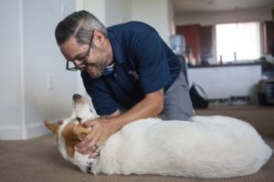
By John Prybys, LAS VEGAS REVIEW-JOURNAL (August 22, 2016) — Randy Dexter and Captain are more than just dog owner and dog. That’s obvious from the way Captain looks for Dexter whenever the Army veteran leaves the room, and the way the Lab mix’s demeanor slips instantly from playful to dead serious once he’s wearing the jacket that denotes his status as a service animal.

Dexter is a retired U.S. Army staff sergeant who did two tours of duty in Iraq. He was diagnosed with both post-traumatic stress disorder and a mild traumatic brain injury, and the story of Dexter and Captain is featured in a new awareness campaign urging veterans and military service people to seek help for traumatic brain injury if they need it.

The campaign, “A Head for the Future,” is sponsored by the Defense and Veterans Brain Injury Center. In his video, Dexter shares the struggles he has experienced coping with his injuries and the reluctance he felt at first to seek help for it.

But, he says, “I was lucky, because when I was in the Army and had my head injury, I was kind of forced to get help.”

Dexter, 34, is a graduate of Green Valley High School who served in the Army for 11 years and had two tours of duty in Iraq. In 2005, Dexter, a combat medic, and his squad were hit by an IED, prompting a long, and continuing, struggle with post-traumatic stress disorder.

Then, after returning home and while still being treated for PTSD and training soldiers bound for Iraq and Afghanistan, Dexter suffered a brain injury during a recreational football game. He’s not sure, even now, what happened. All he knows is that he lost the memory of about 24 hours’ time and, even, of going to the game at all.

X-rays and imaging studies revealed no skull fractures or apparent injuries. But, afterward, Dexter experienced a worsening of already existing problems with his memory, concentration and equilibrium, and began to suffer migraines and severe, debilitating headaches that eventually compounded his PTSD and caused severe depression.

Dr. Scott Livingston, director of education for the Defense and Veterans Brain Injury Center in Silver Springs, Maryland, says symptoms of PTSD and brain injury often can overlap, making diagnosis a challenge. And when a brain injury does occur, he says, it often presents with no obvious symptoms that can be detected by X-ray or imaging scans.

In such cases, the problem likely is “more of a microscopic type of injury within the brain,” Livingston says.

Most civilians probably assume that brain injuries among service people are caused mostly by blasts and blunt-force trauma to the head. Yet, Livingston says, most are caused by motor vehicle collisions, training-related accidents, falls and sports and recreational activities.

Whatever the cause, military personnel are reluctant to report an injury or seek care for it. The current campaign is designed to raise awareness of brain injuries among service people, promote ways to prevent it when possible, and encourage men and women to report it and get it checked out, Livingston says.

“It’s well-known in scientific literature that the earlier someone reports a mild traumatic brain injury and goes to seek help, the better the chances are for better and more complete recovery,” he says.

During his treatment, Dexter participated in a program that paired injured veterans with service dogs. His experience with a dog named Ricochet was so good that he later welcomed the chance to be paired with Captain.

Dexter and Captain are a great team. Dexter says the dog can detect impending anxiety attacks even before he does, and that the dog can serve as a physical shield and protector in such public places as big-box retail stores, which can be particularly unnerving places for those with PTSD.

The true test of Captain’s effectiveness is that the dog has allowed Dexter to significantly reduce the medications he has to take. Today, it would be difficult for someone who doesn’t know the back story to detect Dexter’s struggles with traumatic brain injury, and it was his own previous interest in speaking out publicly about his conditions that led to his participation in the new awareness campaign.

Dexter now attends UNLV, where he’s majoring in communication studies and Spanish. He has been active on the debate teams, will be a peer adviser for other veterans, and hopes to kick off a music show on the university’s HD/internet radio station.

Dexter hopes his video and his story will help to persuade other veterans and active service people to seek out help for PTSD and brain injury. That can be difficult, he notes, because the standard soldier’s stance is that, whatever is happening, “you just deal with it, and that’s true across the whole military culture.”

Read the original article
 

By Alyssa Navarro, Tech Times (August 23, 2016) — Federal health regulators in the United States approved on Monday the use of two new computer softwares as cognitive screening tests for traumatic head injury patients.

Known as ImPACT or the Immediate Post-Concussion Assessment and Cognitive Testing (ImPACT), the new testing device, as well as a similar test designed for children, can be used by doctors to evaluate signs and symptoms of head injuries that could indicate concussion.

ImPACT is designed for patients aged 12 to 59 years old, while ImPACT Pediatric is intended for children aged 5 to 11 years old, officials said. Licensed health care professionals are the only ones allowed to perform the analysis and interpret the results.

The software can be accessed easily because it runs on both desktop computers and laptops, according to the U.S. Food and Drug Administration (FDA). Both tests the first ever devices permitted by the FDA to assess cognitive function after experiencing a possible concussion. They are designed to be part of medical evaluations in hospitals.

Although ImPACT and ImPACT Pediatric will definitely be useful for doctors, both tests are not meant to diagnose concussions or determine treatments that are appropriate for such cases, the FDA said.

Instead, both devices are only designed to test cognitive skills such as reaction time, memory and word recognition. All of these can be impacted by head injuries. Afterwards, the results are compared to a patient’s pre-injury baseline scores or an age-matched control database, the FDA said.

Dr. Carlos Peña, director of the neurological and physical medicine division at the Center for Devices and Radiological Health, acknowledges that the two testing devices can provide useful information that can aid doctors in the evaluation of people who are experiencing potential signs of concussion.

However, Peña says that clinicians should not completely depend on the tests alone to rule out concussion or to decide whether a player with a head injury should return to a game.

Statistics from the Centers for Disease Control and Prevention (CDC) reveal that traumatic brain injuries are responsible for more than 2 million visits to the emergency room in the country annually. Traumatic brain injuries also account for more than 50,000 deaths in America every year.

Cases of head injury among kids have been increasing. In May, a CDC report showed that from January 2001 to December 2013, approximately 214,883 children aged 14 years old and below were brought to emergency departments due to head injuries.

Read the original article
 

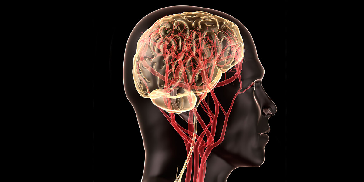
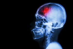
When a person has a stroke, blood flow to the brain is interrupted, causing brain cells to die within minutes due to lack of oxygen. In some cases, this can result in paralysis, speech and language problems, vision problems, and memory loss. But in a new study, researchers have shown that stem cell therapy increases nerve cell production in mice with brain damage due to stroke.

by Marie Ellis, MedicalNewsToday.com (August 22, 2016) — The researchers – led by Berislav Zlokovic, M.D., Ph.D., from the University of Southern California (USC) – publish their findings in the journal Nature Medicine.

According to the Centers for Disease Control and Prevention (CDC), stroke is the fifth leading cause of death in the United States and is also a major cause of disability in adults.

The effects of a stroke depend on the location of the blockage and how much brain tissue is involved, but a stroke on one side of the brain will result in neurological effects on the opposite side of the body.

For example, a stroke on the right side of the brain could produce paralysis on the left side of the body, and vice versa.

A stroke in the brain stem can affect both sides of the body and could leave the patient in a so-called locked-in state, where the patient is unable to speak or move the body below the neck.

Given that about 800,000 people in the U.S. have a stroke each year, the researchers of this latest study wanted to investigate potential therapies.

Therapy is a combination of two methods

The researchers say their therapy is a combination of two methods. One involves surgically grafting human neural stem cells onto the damaged area, where they are able to mature into neurons and other brain cells.

The other therapy uses a compound called 3K3A-APC, which has been shown to help neural stem cells that have been grown in a petri dish grow into neurons. But the researchers say it was not clear what effect the molecule – called activated protein-C (APC) – would have on live animals.

As such, the team used mice for their experiment, and they found that a month after inducing stroke-like brain damage in the mice, those that had received both the stem cells and 3K3A-APC performed much better on motor and sensory function tests, compared with mice that received only one of the treatments or neither.

The researchers also observed that the mice given 3K3A-APC had more stem cells survive and mature into neurons.

But how did the researchers induce stroke-like brain damage in the mice? They disrupted blood flow to a specific brain area.

Then, 1 week later, which is the mouse equivalent of several months in humans, the researchers inserted the stem cells next to the dead tissue and administered either a placebo or 3K3A-APC.

“When you give these mice 3K3A-APC, it works much better than stem cells alone,” says Dr. Zlokovic. “We showed that 3K3A-APC helps the cells convert into neurons and make structural and functional connections with the host’s nervous system.”

‘No one in the stroke field has ever shown this’

The researchers also looked at the connections between the neurons that grew from the stem cells in the damaged brain region and nerve cells in the primary motor cortex.

The team found that the mice given the stem cells and 3K3A-APC had more neuronal connections – synapses – that linked those areas, compared with the mice given the placebo.

Then, when the researchers stimulated the mice’s paws with a vibration, the neurons that grew from the stem cells exhibited a stronger response in the mice that were treated.

“That means the transplanted cells are being functionally integrated into the host’s brain after treatment with 3K3A-APC. No one in the stroke field has ever shown this, so I believe this is going to be the gold standard for future studies.” ~Dr. Berislav Zlokovic

Following on from this study, the researchers want to pursue another phase II clinical trial to examine whether the treatment combination can encourage the growth of new neurons in human stroke patients to improve function.

They say that if that trial is successful, it could be possible to test the therapy’s effects on other conditions, including spinal cord injuries.

“This USC-led animal study could pave the way for a potential breakthrough in how we treat people who have experienced a stroke,” says Jim Koenig, Ph.D., program director at the National Institute of Health’s National Institute of Neurological Disorders and Stroke (NINDS), who funded the study.

“If the therapy works in humans,” he adds, “it could markedly accelerate the recovery of these patients.”

Read the original article

Serving the Brain Injury Community Since 1983