News

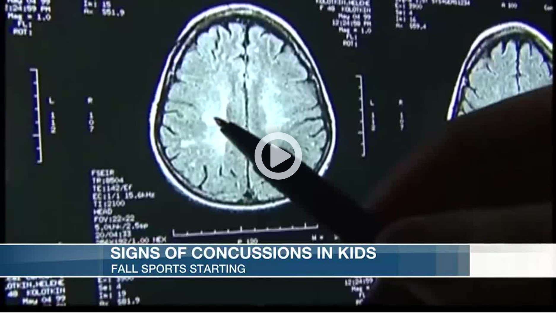
Doctors Discuss Knowing the Signs of Concussion in Young Athletes

By Adria Goins and Alex Onken, KSLA

 
Thousands of students in the Arkansas/Louisiana/Texas (and across the nation) began fall sports over the last few weeks.

However, with the new season here, comes a risk of injury. Football is the leading sport when it comes to concussions.

The signs of a concussion are headache, fatigue and nausea. Parents are advised to then bring their child to a doctor right away if suspecting a possible concussion.

“First diagnose it early and then after you diagnose it early make sure you avoid the triggers. So avoid extra screen time, over-exercising and just basically have 24 to 48 hours of cognitive physical rest,” said Dr. Kenneth Aguirre of Oschner-LSU Health Shreveport, who specializes in sports medicine.

According to Dr. Charles Webb, also with Oschner-LSU Health and a sports medicine specialist, the topic of concussions and the potential risks of football comes up often.

“I get that question a lot from parents. They want to know is it safe for my child to play high school or junior high, or even pee wee or popcorn football. and the question comes up because parents are worried about concussions. So my answer to them is if it were my child I wouldn’t let them play until they had an organized professional coach teaching them both how to hit and receive a hit.”

Young athletes are usually taught how to hit and receive a hit around junior high. Dr. Webb said parents should put their children in club soccer or flag football in contrast to popcorn or pee wee football.

“It’s much safer and you’re less likely to get hit in the head,” he said. “And you still get all the conditioning you need to play football later on in life.”

In addition, doctors say keeping children awake when they have a concussion is a common misconception. Sleep is actually very good for the healing process.
 

CLICK HERE to read the original article


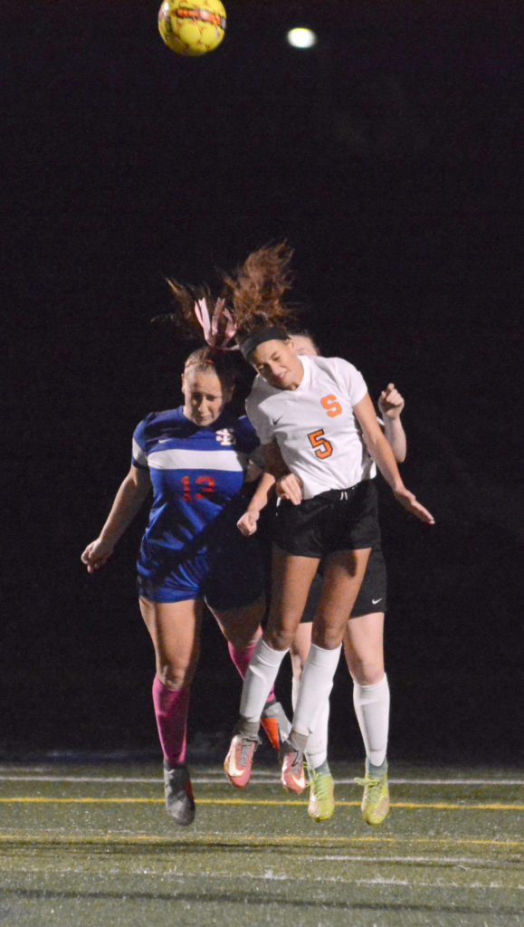
High school injury reports analyzed by InvestigateWest and Pamplin Media show that girls are twice as likely to get concussions as boys in Oregon. Girls in the 13U age group, pictured above, are the youngest allowed to use headers.
 

The Concussion Gap: Head injuries in girls soccer are an ‘Unpublicized Epidemic’

Lee van der Voo, InvestigateWest, photos by David Ball / Pamplin Media Group

 
When it comes to concussion in sports, all eyes are on football, or so it seems. But it’s not just football that causes a high number of head injuries among young athletes.

Another culprit? Girls soccer.

National research has found girls are more likely to suffer a concussion than boys in any sport. In 2017, researchers at Northwestern University generated national headlines when they found concussion rates among young female soccer players were nearly as high as concussion rates for boys playing football — and roughly triple the rate of concussions in boys soccer.

In Oregon, injury reports from public high schools analyzed by InvestigateWest and Pamplin Media Group mirrored that trend, showing soccer concussions were second to those from football between 2015 and 2017. What’s more, at the schools that included the gender of injured athletes, there were nearly twice as many reports of possible concussions for girls playing soccer than boys in the sport.

The rate of concussions in girls soccer worries local experts like Jim Chesnutt, a doctor in sports medicine at Oregon Health & Science University, who says those injuries are not widely recognized, even as concussion rates rise for girls playing soccer.

“In a lot of ways, it’s a growing epidemic for young girls that I think has gone unpublicized,” said Chesnutt, co-director of the Oregon Concussion Awareness and Management Program and a member of the Governor’s Task Force on Traumatic Brain Injury.

More exposure, more injury

It’s understandable that much of the youth concussion conversation centers on football, given the physical contact that is visibly — and audibly — evident on every play, as well as the large rosters and the lengthy lists of players who are injured.

But if you compare girls soccer with football, and only look at the high school participation and injury data, “you’re missing a gigantic part of the picture,” according to Michael Koester, a doctor of sports medicine at the Slocum Center in Eugene. He directs its sports concussion program and serves as the chair of the Sports Medicine Advisory Committee for the National Federation of State High School Associations.

Koester notes that high school boys play eight to 10 football games per season, and typically play other sports in the off-season.

Girls, however, play 15 to 20 soccer games in a high school season, but when that season ends, they may play another 80-plus games throughout the winter, spring and summer with club teams, said Koester, who, like Chesnutt, is a medical adviser to the Oregon Schools Activities Association.

“If we’re looking at injury risk by athletic exposure,” which is one practice or game, a standard in evaluating risk, Koester said, female soccer players probably are playing five if not 10 times more practices and games than football players.

And Koester doesn’t see the trend ending.

“The thought used to be that this was all revolving around, ‘Wow! They want to get their kid a scholarship,’ ” he said. “Now it’s kind of gotten to the point where there’s so much single-sport participation that we see kids that are specializing in sport early, just so they’ll be able to make their high school team.”

Single-sport athletes are more prone to injury in any sport. According to a study by scientists at the University of Wisconsin, high school athletes who specialized in just one sport at an early age were twice as likely to suffer injuries to their lower extremities.

“We see a lot of overuse injury among girls playing soccer,” Koester said. “We see a lot of ACL injury among girls playing soccer. It’s a well-known problem.”

Aggressive play

Another factor is the evolution of sports.

Angella Bond is an athletic trainer for Tuality Sports Medicine and works on the sidelines with athletes at Hillsboro schools. Anecdotally, she said, all athletes push to be bigger, faster and stronger. Soccer is no exception, nor are girls.

As athletes develop, they take bigger hits at higher speeds, and competitive games build on their momentum. As competition grows in girls soccer, the sport is trending to be more aggressive, she said.

“Unfortunately, I think that happens with girls sports,” she said. “Arms fly a little bit more.”

Chesnutt agreed. “I think over the years, soccer has become more physical,” he said. “And I think the physical contact and the aggressive nature of that physical contact is more associated with concussions.”

According to the American Academy of Pediatrics, soccer — unlike football, ice hockey and lacrosse — is not a “collision sport.” But it is a “contact sport” because athletes “routinely make contact with each other or inanimate objects.”

Header balls, though often singled out as a source of concussions, are not necessarily to blame.

The force created when a soccer ball meets a head can rattle a brain, but data increasingly points to other factors when competitors vie for a ball in the air.

According to a study by The Research Institute at Nationwide Children’s Hospital, while headers accounted for 27 percent of concussions, it was knocks with other players on aerial play — including head-to-head contact and arms and elbows to the head — and contact with the ground that accounted for 70 percent of those concussions in girls soccer, suggesting aggressive play is a factor in most concussions involving headers.

Why girls?

But why are girls more prone to concussions than boys while playing soccer? The prevailing theories focus on their weaker neck-muscle development, weaker body strength (needed to stabilize the neck and head during aerial play), and more frequent contact with the ground. A year ago, a study in the Journal of the American Osteopathic Association found that female high school soccer players took twice as long as male players to recover.

It’s also possible that girls don’t benefit as much from early treatment. A recent study published by the American Academy of Pediatrics found that girls are five times more likely than boys to stay on the pitch and play through a head injury.

And the soccer community has been slow to recognize the hard hits its girls are taking. Instead, soccer is at the forefront of the cultural empowerment of girls.

Local experts concerned about concussion risk note that sports, including girls soccer, have plenty of benefits. Just being physically active is good for kids, and sports like soccer help establish lifelong fitness habits, teach team-building skills, and promote character development and assertiveness.

“The worry is that the take-home message is that (girls soccer) is healthy and fantastic and nothing can be bad about it,” said Koester, who says an opposite negative message, equally extreme, is more often associated with boys playing football.

Greater awareness needed

Concussion education and awareness in girls soccer is paramount, according to local experts such as Chesnutt.

“I think the way to decrease it is to really analyze how we can modify the amount of body contact that goes on in soccer to limit the dangerous aggressive behavior that is associated with concussion,” he said.

Unlike youth football, a sport that’s adjusting to new information about concussions all the time, soccer has largely failed to address new information about concussions, Chesnutt said.

Football, for example, has reduced head-to-head helmet play, limited full-contact practices and games, and zeroed in on the specialty teams with the highest concussion rates.

“Football has really done, I think, an exceptional job of identifying some areas where there have been some definite higher incidents and some problems,” said Chesnutt, who lectures nationally about youth concussions. “As a group of coaches, leagues, parents and referees, they’ve all looked at it and come up with some solutions that have decreased concussion rates. And I think it’s time for soccer to do the same thing.”

Read the original article


New Rules to Protect Your Kid’s Noggin

May 25, 2019, Parents Magazine

 
Children bonk their head all the time when they’re wrestling with siblings, playing soccer, and just being clumsy-and it’s easy to worry that a bump could turn into something bigger. After all, more than 800,000 kids in the U.S. get a concussion every year. For the first time, the Centers for Disease Control and Prevention has released specific “return to learn” and “return to play” guidelines for head injuries, based on 25 years of research. One doctor shares the big takeaways.

ALWAYS take any injury beyond a light head bump seiously. A concussion occurs when a bump, blow, or jolt to the head or a hit to the body makes the brain bounce or twist in the skull. This creates chemical changes and can sometimes damage brain cells. “If your child complains of a headache or dizziness, is nauseous or vomiting, appears dazed, or sleeps more or less than usual, it’s time to get a doctor’s evaluation,” says Dennis Cardone, D.O., associate professor of orthopedic surgery and pediatrics and co-director of the NYU Langone Concussion Center. Even toddlers can get a concussion from a tumble, so look for changes in their behavior such as not wanting to nurse or eat or losing interest in toys.

If diagnosed with a concussion, your child will need menlal rest, says Dr. Cardone. That means taking a break from all activities for two to three days, and after that, starting with light aerobic activity. He may need to attend school for only half the day or do little to no homework (he won’t mind this rule!). However, he shouldn’t return to any sports or strenuous activities that have a high risk of falling or contact (think: field hockey, gymnastics, climbing a tree) until he’s been cleared by his doctor, which should be within a few weeks.

Download the original article PDF
 

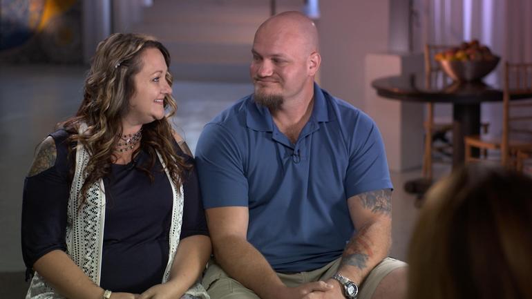
Libby and Tom Bates // CBS News

A brain disease best known for impacting football players who suffered concussions is now being found in soldiers

By Sharyn Alfonsi, September 16, 2018, CBS News

Until a few years ago, NFL players who struggled with severe depression, bouts of rage and memory loss in their retirement were often told they were just having a hard time adjusting to life away from the game. Doctors have since learned these changes can be symptoms of the degenerative brain disease CTE – chronic traumatic encephalopathy, caused by blows to the head.

As we first reported in January, CTE isn’t just affecting athletes, but also showing up in our nation’s heroes. Since 9/11 over 300,000 soldiers have returned home with brain injuries. Researchers fear the impact of CTE could cripple a generation of warriors.

When Joy Kieffer buried her 34-year old son this past summer, it was the end of a long goodbye.

Kieffer’s son, Sgt. Kevin Ash, enlisted in the Army Reserves at the age of 18. Over three deployments, he was exposed to 12 combat blasts, many of them roadside bombs. He returned home in 2012 a different man.

Joy Kieffer: His whole personality had changed. I thought it was exposure to all of the things that he had seen, and he had just become harder. You know, but he was — he was not happy.

Sharyn Alfonsi: So at this point, you’re thinking this decline, this change in my child is just that he’s been in war and he’s seen too much.

Joy Kieffer: Right.

Sharyn Alfonsi: Did he tell you about blasts that he experienced during that time?

Joy Kieffer: Uh-huh.

Sharyn Alfonsi: What did he–tell you?

Joy Kieffer: That they shook him. And he was having blackouts. And — it frightened him.

Ash withdrew from family and friends. He was angry. Depressed. Doctors prescribed therapy and medication, but his health began to decline quickly. By his 34th birthday, Sgt. Kevin Ash was unable to speak, walk or eat on his own.

Sharyn Alfonsi: Looking back on it now, was there anything you feel like he could’ve done?

Joy Kieffer: Uh-uh.

Sharyn Alfonsi: Because?

Joy Kieffer: Because it was– it– it was his brain. The thing I didn’t know was that his brain was continuing to die. I mean, before he went into the service he said, “you know, I could come back with no legs, or no arms, or even blind, or I could be shot, I could die,” but nobody ever said that he could lose his mind one day at a time.

His final wish was to serve his country one last time by donating his brain to science — a gesture he thought would bring better understanding to the invisible wounds of war.

Joy reached out to the VA-Boston University-Concussion Legacy Foundation Brain Bank where neuropathologist Dr. Ann McKee is leading the charge in researching head trauma and the degenerative brain disease CTE.

McKee has spent fourteen years looking at the postmortem brains of hundreds of athletes who suffered concussions while playing their sport.

Last summer, her findings shook the football world when she discovered CTE in the brains of 110 out of 111 deceased NFL players — raising serious concerns for those in the game today.

And when Dr. McKee autopsied Patriots tight-end Aaron Hernandez who killed himself after being convicted of murder, she found the most severe case of CTE ever, in someone under 30.

Now she’s seeing similar patterns in deceased veterans who experienced a different kind of head trauma — combat blasts. Of the 125 veterans’ brains Dr. Mckee’s examined, 74 had CTE.

Sharyn Alfonsi: I can understand a football player who keeps, you know, hitting his head, and having impact and concussions. But how is it that a combat veteran, who maybe just experienced a blast, has the same type of injury?

Dr. Ann McKee: This blast injury causes a tremendous sort of– ricochet or– or– a whiplash injury to the brain inside the skull and that’s what gives rise to the same changes that we see in football players, as in military veterans.

Blast trauma was first recognized back in World War I. Known as ‘shell shock,’ poorly protected soldiers often died immediately or went on to suffer physical and psychological symptoms. Today, sophisticated armor allows more soldiers to walk away from an explosion but exposure can still damage the brain — an injury that can worsen over time.

Dr. Ann McKee: It’s not a new injury. But what’s been really stumping us, I think, as– as physicians is it’s not easily detectable, right? It’s– you’ve got a lot of psychiatric symptoms– and you can’t see it very well on images of the brain and so it didn’t occur to us. And I think that’s been the gap, really, that this has been what everyone calls an invisible injury.

Dr. Ann McKee: This is the world’s largest CTE brain bank.

The only foolproof way to diagnose CTE is by testing a post-mortem brain.

Sharyn Alfonsi: So these are full of hundreds of brains…

Dr. Ann McKee: Hundreds of brains, thousands really…

Researchers carefully dissect sections of the brain where they look for changes in the folds of the frontal lobes – an area responsible for memory, judgement, emotions, impulse control and personality.

Dr. Ann McKee: Do you see there’s a tiny little hole there? That is an abnormality. And it’s a clear abnormality.

Sharyn Alfonsi: And what would that affect?

Dr. Ann McKee: Well, it’s part of the memory circuit. You can see that clear hole there that shouldn’t be there. It’s connecting the important memory regions of the brain with other regions. So that is a sign of CTE.

Thin slivers of the affected areas are then stained and viewed microscopically. It’s in these final stages where a diagnosis becomes clear as in the case of Sgt. Kevin Ash.

Sharyn Alfonsi: So this is Sergeant Ash’s brain?

Dr. Ann McKee: Right. This is– four sections of his brain. And what you can see is– these lesions. The, and those lesions are CTE And they’re in very characteristic parts of the brain. They’re at the bottom of the crevice. That’s a unique feature of CTE.

Sharyn Alfonsi: And in a healthy brain, you wouldn’t see any of those kind of brown spots?

Dr. Ann McKee: No, no, it would be completely clear. And then when you look microscopically, you can see that the tau, which is staining brown and is inside nerve cells is surrounding these little vessels.

Sharyn Alfonsi: And explain, what is the tau?

Dr. Ann McKee: So tau is a protein that’s normally in the nerve cell. It helps with structure and after trauma, it starts clumping up as a toxin inside the nerve cell. And over time, and even years, gradually that nerve cell dies.

Dr. Lee Goldstein has been building on Dr. McKee’s work with testing on mice.

Inside his Boston University lab, Dr. Goldstein built a 27-foot blast tube where a mouse – and in this demonstration, a model – is exposed to an explosion equivalent to the IEDs used in Iraq and Afghanistan.

Dr. Lee Goldstein: When it reaches about 25 this thing is going to go.

Dr. Goldstein’s model shows what’s going on inside the brain during a blast. The brightly colored waves illustrate stress on the soft tissues of the brain as it ricochets back and forth within the skull.

Dr. Lee Goldstein: What we see after these blast exposures, the animals actually look fine. Which is shocking to us. So they come out of what is a near lethal blast exposure, just like our military service men and women do. And they appear to be fine. But what we know is that that brain is not the same after that exposure as it was microseconds before. And if there is a subsequent exposure, that change will be accelerated. And ultimately, this triggers a neurodegenerative disease. And, in fact, we can see that really after even one of these exposures.

Sharyn Alfonsi: The Department of Defense estimates hundreds of thousands of soldiers have experienced a blast like this. What does that tell you?

Dr. Lee Goldstein: This is a disease and a problem that we’re going to be dealing with for decades. And it’s a huge public health problem. It’s a huge problem for the Veterans Administration. It’s a huge moral responsibility for all of us.

A responsibility owed to soldiers like 34-year-old Sgt. Tom Bates.

Sgt. Tom Bates: We were struck with a large IED. It was a total devastation strike.

Bates miraculously walked away from a mangled humvee — one of four IED blasts he survived during deployments in Iraq and Afghanistan.

Sharyn Alfonsi: Do you remember feeling the impact in your body?

Sgt. Tom Bates: Yes. Yeah.

Sharyn Alfonsi: What does that feel like?

Sgt. Tom Bates: Just basically like getting hit by a train.

Sharyn Alfonsi: And you were put back on the frontlines.

Sgt. Tom Bates: Yes.

Sharyn Alfonsi: And that was it?

Sgt. Tom Bates: Uh-huh

When Bates returned home in 2009, his wife Libby immediately saw a dramatic change.

Libby Bates: I thought, “Something is not absolutely right here. Something’s going on. For him to just lay there and to sob and be so sad. You know, what do you do for that? How do I– how do I help him? He would look at me and say, “If it wasn’t for you, I would end it all right now.” You know, I mean, like, what do you– what do you do– and what do you say to somebody who says that? You know I love this man so much. And —

Sharyn Alfonsi: You’re going to the VA, you’re getting help, but did you feel like you weren’t getting answers?

Sgt. Tom Bates: Yes.

Sharyn Alfonsi: And so you took it into your own hands and started researching?

Sgt. Tom Bates: I knew the way everything had gone and how quick a lot of my neurological issues had progressed that something was wrong. And I just– I wanted answers for it.

That led him to New York’s Mount Sinai Hospital where neurologist Dr. Sam Gandy is trying to move beyond diagnosing CTE only in the dead by using scans that test for the disease in the living.

Dr. Sam Gandy: By having this during life, this now gives us for the first time the possibility of estimating the true prevalence of the disease. It’s important to estimate prevalence so that people can have some sense of what the risk is.

In the past year, 50 veterans and athletes have been tested for the disease here. Tom Bates asked to be a part of it.

That radioactive tracer – known as t807 – clings to those dead clusters of protein known as tau, which are typical markers of the disease.

Through the course of a 20 minute PET scan, high resolution images are taken of the brain and then combined with MRI results to get a 360 degree picture of whether there are potential signs of CTE.

Scan results confirmed what Tom and Libby had long suspected.

On the right, we see a normal brain scan with no signs of CTE next to Tom’s brain where tau deposits, possible markers of CTE, are bright orange.

Dr. Sam Gandy: Here these could be responsible for some of the anxiety and depression he’s suffered and we’re concerned it will progress.

Sgt. Tom Bates: My hope is that this study becomes more prominent, and gets to more veterans, and stuff like that so we can actually get, like, a reflection of what population might actually have this.

There is no cure for CTE.

Dr. Gandy hopes his trial will lead to drug therapies so he can offer some relief to patients like Tom.

Dr. Ann McKee believes some people may be at higher risk of getting the disease than others.

While examining NFL star Aaron Hernandez’s brain she identified a genetic bio-marker she believes may have predisposed him to CTE.

A discovery that could have far-reaching implications on the football field and battlefield.

Sharyn Alfonsi: Do you think you will ever be your old self again?

Sgt. Tom Bates: I don’t ever see me being my old self again. I think it’s just too far gone.

Sharyn Alfonsi: So what’s your hope then?

Sgt. Tom Bates: Just to not become worse than I am now.

Since our story first aired, over 100 veterans have contacted Dr. Gandy to enroll in ongoing trials to identify whether they are living with CTE. And more than 300 have reached out to Dr. Mckee about donating their brains to research.

Read the original article
 

Junior Seau, shown at his beloved Pacific Ocean in the ESPN Films “30 for 30” documentary “Seau,” which premieres Thursday. (ESPN Films)

ESPN hits the mark with documentary ‘Seau’

By Tom Krasovic, September 20, 2018, San Diego Union Tribune

An aerial view of the Oceanside coast, in full sparkle and splendor below, grandly eases viewers into “Seau,” an ESPN Films documentary in the “30 for 30” series that debuts Thursday on the streaming service ESPN+.

It’s a sunny scene, the Pacific Ocean’s turquoise waves illuminated as they roll toward the white beach. The late Junior Seau told friends he found peace paddling on these waters, deep into his life alongside the town where he’d grown up.

Up at dawn with a yellow long-board and oar in hand, Seau had only a short walk from his beachfront home to the water.

Yet the former Chargers linebacker, role model and local philanthropist was then also writing in a journal of bouts with depression, memory loss and perceived guilt. There were headaches, too, and nights plagued by insomnia. “Buddy,” he’d told a friend and professional soccer player who’d suffered a brain injury from heading a ball, “I’ve had a concussion since I was 15.”

Diary entries also revealed feelings of humiliation and embarrassment over not living up to expectations of others and himself, and of feeling used by others.

“The world has nothing for me,” Seau pens in one entry, the cursive words all too legible.

One of Seau’s surviving adult children, after reading the grim line aloud, wonders why his father didn’t regard his family as something in this apparent world of nothing.

Why couldn’t they have been a lifeline for him to reach out and grasp?

“Seau,” produced and directed by Kirby Bradley, lets viewers draw their own conclusions about a complicated life that ended one May morning six years ago, at age 43, with a self-inflicted gunshot wound to the chest, but not before we hear from an array of family members, friends and experts in football and brain science.

At the end of the 90-minute film, themes of redemption and hope are raised.

“Let’s all walk from here being better for having known Junior Seau and the impact he had on our lives,” NFL quarterback Drew Brees, a former Chargers teammate of the Hall of Fame linebacker, concludes near the film’s end.

Former Chargers lineman Aaron Taylor notes that in death, Seau drew extraordinary attention to the link between head trauma and a degenerative brain disease, CTE, revealed in a tissue sample sent to a brain scientists at the family’s request.

Exciting beginnings and success are a thread to the film, followed often by bitter detours or hurtful endings.

Seau took to sports at Oceanside High with a passion that rivaled his stunning blend of size, speed and agility. If he was slamming into football ball-carriers or catching passes, scoring baskets or throwing the discus and shot, he was a “force of nature” for the green-and-white-clad Pirates, observers said.

A flood of football scholarship offers came to the small home where Seau and his brothers slept in a tiny garage.

Jubilation ensued when Seau chose USC, keeping him close to his parents and siblings and the tight-knit Samoan-American community in Oceanside. A similar celebration arose in 1990 when the Chargers drafted him fifth overall. “I’m a real momma’s boy,” Seau said, pulling on a blue team cap.

Playing for his beloved “Diego,” he led the long-struggling Chargers to the playoffs in just his third season, and their first Super Bowl two years later. “Now the world is gonna know the San Diego Chargers,” he told some 70,000 celebrants in Mission Valley after the team returned from claiming the 1994 AFC title in Pittsburgh.

The flip side?

If Oceanside lost a game in which he played, Junior lost his lunch money. It was the price his father exacted.

The thrill of signing with USC gave way to humiliation when a failed admittance test made him ineligible as a freshman. His father refused to talk to him in response, deeming the failure an embarrassment to the family. After a dominant junior year with USC, there would be no senior year. Making money was the next step, in no small part because he wanted to support his parents and other family members.

The Chargers couldn’t build upon their Super Bowl season, and the team’s constant losing wore on Seau.

When the Chargers traded him in the spring of 2003, after 13 seasons with the club, Seau was hurt that the team — Stay Unclassy, San Diego? — called not him but his agent to tell him the news. “I know that was hard on him,” said the agent, Steve Feldman.

Gina Seau was working for the Chargers in marketing when she first met Seau early in his NFL career.

She recalled “very kind eyes” and a “very soft voice” that almost “didn’t match the size and stature.”

The two would marry, but erratic behavior that Gina Seau linked to numerous football-related head injuries — “My head is on fire,” he told her — led to a divorce in 2002. The two remained friends. Believing that driving off a steep coastal cliff in October 2010 wasn’t an accident, Gina pleaded with her former husband to get help.

Here’s hoping that if there’s a “Seau II,” events yet to transpire bring more developments of redemption. Say, a cure for CTE.

Read the original article


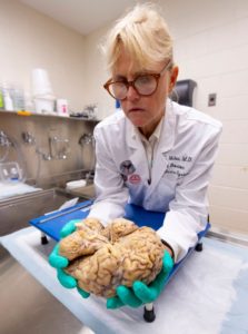
Ann C. McKee, chief of neuropathology at the VA Boston Healthcare System, which houses the world’s largest brain bank devoted to CTE research, examines a brain earlier this month.(Photo: Robert Deutsch, USA TODAY)

Researchers close in on CTE diagnosis in living, one brain at a time

By Nancy Armour, August 24, 2018, USA TODAY

BOSTON – Submerged in chemicals in the stainless-steel bowl is the key to life and, researchers hope, death.

It’s a human brain. That of a man who played college football in the 1950s, to be exact. His family donated his brain to get answers for themselves, but what’s found could lead to more answers about chronic traumatic encephalopathy, the devastating neurodegenerative disease linked to concussions and repetitive head trauma from football and other contact sports.

“Our main objective, our overarching goal, is to help the people who are living. To be able to diagnose this disease during life,” says Ann McKee, chief of neuropathology at the VA Boston Healthcare System, which houses the world’s largest brain bank devoted to CTE research.

“If we can diagnose it, we can monitor it and test therapies to see if they’re effective in treating this disease,” says McKee, director of the CTE Center at Boston University’s School of Medicine. “It would really dramatically increase our ability to point out genetic susceptibilities for this. We’d be able to look at how much is too much in certain individuals or certain positions in certain sports.”

As another football season begins, it inevitably leads to questions and fears about head trauma and its long-term damage. How many hits are too many? What can parents do to protect their children or players do to protect themselves? Are athletes in certain sports more susceptible?

Most important, which athletes will develop CTE – or Parkinson’s or ALS (amyotrophic lateral sclerosis) – and why?

The answers will come from brains such as the one McKee dissected this month, when USA TODAY Sports toured the brain bank.

The brain bank has more than 500 brains, most of them donated by former athletes or their families who suspected CTE because of mood swings, behavioral changes, depression or dementia. Of those brains, more than 360 had CTE, McKee says.

SEARCHING FOR CLUES

The arrival of a brain sets two teams in motion. One set of clinicians talks to the family to find out more about the donors. Did they play any sports? If so, what and for how long? When did they start? Did they experience any other kind of head trauma, say from an automobile accident, domestic violence or military service? Did they have drug or alcohol problems? How did their mental health change, and when did that occur?

Separately, and usually without any information about the person whose brain it was, McKee and her researchers study the brain. It is cut in half, and one half is stored in a minus-80-degree freezer, so it will be available for molecular, genetic and biochemical studies.

The other half is then photographed and sectioned. After removing the brain stem, McKee uses what looks like a bread knife to cut slices of the brain about a quarter-inch thick.


Ann C. McKee slices the brain into segments about a quarter-inch thick as part of in-depth, time consuming research on the organ. McKee hopes the work will unlock answers to CTE. (Photo: Robert Deutsch, USA TODAY)
 
Simply by looking at the brain, McKee can tell a few things. The brain of this man, who was in his 80s when he died, has shrunk, noticeably smaller than it should be for a man who once played football. The folds of the brain, normally pressed tightly against one another, are loose and have gaps between them, some large enough that the tip of a finger could be inserted.

She points to the ventricles, chambers in the middle of his brain that are filled with fluid during life. They should be small, but these are “just gigantic.”

“As the brain shrinks, they expand. What this indicates is there’s been enormous shrinkage of the brain,” McKee says. “Those are huge.”

The hippocampus, a section in the middle of the brain that controls memory, is small but not abnormally so for a man in his 80s. If it was, that could be an indication of Alzheimer’s. But a membrane that runs from one side of the brain to the other, normally thick like a rubber band, has shrunk. In some spots, it’s almost invisible.

“This is looking more like frontal predominant atrophy, and that could mean CTE because Alzheimer’s almost always affects the hippocampus,” McKee says. “At this point, I always want to know, ‘What is it? Let’s look under the microscope.’ But you have to wait.”

CTE can’t be seen by the naked eye, and it takes at least three weeks to prepare slides of the brain tissue.


CTE is caused by tau, a protein in the brain released as a result of head trauma. When tau clumps together, it damages brain cells and can change the brain’s function. Though tau causes Alzheimer’s, McKee says, the tau that causes CTE looks distinctly different.

Under a microscope, it can be seen in telltale brown spots.

“CTE is very focal. In fact, in its early stages, it’s in the crevices. It just piles up. And that’s around blood vessels,” McKee says. “That’s very different. Alzheimer’s never does that.”

As CTE progresses, those clusters or clumps of tau will spread, and the disease will become more severe. That’s why, in the early stages of disease, stages 1 and 2, the symptoms usually relate to behavioral changes or mood swings. In stages 3 and 4, the disease is exhibited in memory loss.

“We think there may be more pathology in the young players than we’re appreciating just with the tau protein,” McKee says. “We think there’s maybe white matter structural changes or maybe inflammatory changes that are responsible for that loss of control, which is so difficult for the individuals.”

‘EVERY CASE IS A MYSTERY’

Once the slides have been examined, the pathologists and clinicians will come together for a conference. At this point, neither knows what the other does. The clinicians detail what they’ve learned about the brain donor’s history and suggest a diagnosis. The pathologists will then say whether the brain tissue confirms it.

“Every case is a mystery,” McKee says. “It’s not the same way you usually solve a mystery. I solve the pathology first, and then you go back and find out (the history). And then you try and put the two together.”

Some former players and their families once were reluctant to donate their brains, but that stigma largely has disappeared. So much so that McKee said brains arrive at the Boston bank almost every day.

Though that lengthens the time it takes to reach a definitive diagnosis, it will shorten the time before a living diagnosis can be found. In addition to the work done in her lab, McKee shares tissue samples with researchers around the world.

“What we want to do is establish the risk, educate people, educate parents, educate players,” McKee says. “So if they’re unwilling to risk that future self, if they’re unwilling to take that risk because it’s too high for them personally, we want to give them enough data so they can make a very sound and wise decision.”

When that day comes, it will change sports forever.

Read the original article
 

By John Prybys, LAS VEGAS REVIEW-JOURNAL (August 22, 2016) — Randy Dexter and Captain are more than just dog owner and dog. That’s obvious from the way Captain looks for Dexter whenever the Army veteran leaves the room, and the way the Lab mix’s demeanor slips instantly from playful to dead serious once he’s wearing the jacket that denotes his status as a service animal.

Dexter is a retired U.S. Army staff sergeant who did two tours of duty in Iraq. He was diagnosed with both post-traumatic stress disorder and a mild traumatic brain injury, and the story of Dexter and Captain is featured in a new awareness campaign urging veterans and military service people to seek help for traumatic brain injury if they need it.

The campaign, “A Head for the Future,” is sponsored by the Defense and Veterans Brain Injury Center. In his video, Dexter shares the struggles he has experienced coping with his injuries and the reluctance he felt at first to seek help for it.

But, he says, “I was lucky, because when I was in the Army and had my head injury, I was kind of forced to get help.”

Dexter, 34, is a graduate of Green Valley High School who served in the Army for 11 years and had two tours of duty in Iraq. In 2005, Dexter, a combat medic, and his squad were hit by an IED, prompting a long, and continuing, struggle with post-traumatic stress disorder.

Then, after returning home and while still being treated for PTSD and training soldiers bound for Iraq and Afghanistan, Dexter suffered a brain injury during a recreational football game. He’s not sure, even now, what happened. All he knows is that he lost the memory of about 24 hours’ time and, even, of going to the game at all.

X-rays and imaging studies revealed no skull fractures or apparent injuries. But, afterward, Dexter experienced a worsening of already existing problems with his memory, concentration and equilibrium, and began to suffer migraines and severe, debilitating headaches that eventually compounded his PTSD and caused severe depression.

Dr. Scott Livingston, director of education for the Defense and Veterans Brain Injury Center in Silver Springs, Maryland, says symptoms of PTSD and brain injury often can overlap, making diagnosis a challenge. And when a brain injury does occur, he says, it often presents with no obvious symptoms that can be detected by X-ray or imaging scans.

In such cases, the problem likely is “more of a microscopic type of injury within the brain,” Livingston says.

Most civilians probably assume that brain injuries among service people are caused mostly by blasts and blunt-force trauma to the head. Yet, Livingston says, most are caused by motor vehicle collisions, training-related accidents, falls and sports and recreational activities.

Whatever the cause, military personnel are reluctant to report an injury or seek care for it. The current campaign is designed to raise awareness of brain injuries among service people, promote ways to prevent it when possible, and encourage men and women to report it and get it checked out, Livingston says.

“It’s well-known in scientific literature that the earlier someone reports a mild traumatic brain injury and goes to seek help, the better the chances are for better and more complete recovery,” he says.

During his treatment, Dexter participated in a program that paired injured veterans with service dogs. His experience with a dog named Ricochet was so good that he later welcomed the chance to be paired with Captain.

Dexter and Captain are a great team. Dexter says the dog can detect impending anxiety attacks even before he does, and that the dog can serve as a physical shield and protector in such public places as big-box retail stores, which can be particularly unnerving places for those with PTSD.

The true test of Captain’s effectiveness is that the dog has allowed Dexter to significantly reduce the medications he has to take. Today, it would be difficult for someone who doesn’t know the back story to detect Dexter’s struggles with traumatic brain injury, and it was his own previous interest in speaking out publicly about his conditions that led to his participation in the new awareness campaign.

Dexter now attends UNLV, where he’s majoring in communication studies and Spanish. He has been active on the debate teams, will be a peer adviser for other veterans, and hopes to kick off a music show on the university’s HD/internet radio station.

Dexter hopes his video and his story will help to persuade other veterans and active service people to seek out help for PTSD and brain injury. That can be difficult, he notes, because the standard soldier’s stance is that, whatever is happening, “you just deal with it, and that’s true across the whole military culture.”

Read the original article
 

By Alyssa Navarro, Tech Times (August 23, 2016) — Federal health regulators in the United States approved on Monday the use of two new computer softwares as cognitive screening tests for traumatic head injury patients.

Known as ImPACT or the Immediate Post-Concussion Assessment and Cognitive Testing (ImPACT), the new testing device, as well as a similar test designed for children, can be used by doctors to evaluate signs and symptoms of head injuries that could indicate concussion.

ImPACT is designed for patients aged 12 to 59 years old, while ImPACT Pediatric is intended for children aged 5 to 11 years old, officials said. Licensed health care professionals are the only ones allowed to perform the analysis and interpret the results.

The software can be accessed easily because it runs on both desktop computers and laptops, according to the U.S. Food and Drug Administration (FDA). Both tests the first ever devices permitted by the FDA to assess cognitive function after experiencing a possible concussion. They are designed to be part of medical evaluations in hospitals.

Although ImPACT and ImPACT Pediatric will definitely be useful for doctors, both tests are not meant to diagnose concussions or determine treatments that are appropriate for such cases, the FDA said.

Instead, both devices are only designed to test cognitive skills such as reaction time, memory and word recognition. All of these can be impacted by head injuries. Afterwards, the results are compared to a patient’s pre-injury baseline scores or an age-matched control database, the FDA said.

Dr. Carlos Peña, director of the neurological and physical medicine division at the Center for Devices and Radiological Health, acknowledges that the two testing devices can provide useful information that can aid doctors in the evaluation of people who are experiencing potential signs of concussion.

However, Peña says that clinicians should not completely depend on the tests alone to rule out concussion or to decide whether a player with a head injury should return to a game.

Statistics from the Centers for Disease Control and Prevention (CDC) reveal that traumatic brain injuries are responsible for more than 2 million visits to the emergency room in the country annually. Traumatic brain injuries also account for more than 50,000 deaths in America every year.

Cases of head injury among kids have been increasing. In May, a CDC report showed that from January 2001 to December 2013, approximately 214,883 children aged 14 years old and below were brought to emergency departments due to head injuries.

Read the original article
 

UCSF Researchers Advocate Prioritizing Teens for Education and Prevention

by Scott Maier (August 17, 2016) — The number of Americans diagnosed with concussions is growing, most significantly in adolescents, according to researchers at UC San Francisco. They recommend that adolescents be prioritized for ongoing work in concussion education, diagnosis, treatment and prevention.

The findings appear online August 16, 2016, in the Orthopaedic Journal of Sports Medicine.

“Our study evaluated a large cross-section of the U.S. population,” said lead author Alan Zhang, MD, UCSF Health orthopaedic surgeon. “We were surprised to see that the increase in concussion cases over the past few years mainly were from adolescent patients aged 10 to 19.”

Concussions are a form of mild traumatic brain injury resulting in transient functional and biochemical changes in the brain. They can lead to time lost from sports, work and school, as well as significant medical costs.

Though symptoms resolve in most concussion patients within weeks, some patients’ symptoms last for months, including depression, headache, dizziness and fogginess. Neuroimaging and neuropathological studies also suggest there may be chronic structural abnormalities in the brain following multiple concussions.

Recent studies have shown an increase in traumatic brain injuries diagnosed in many U.S. emergency departments. Smaller cohort studies of pediatric and high school athletes also have indicated a rise in concussions for certain sports, such as football and girls’ soccer. However, this is the first study to assess trends in concussion diagnoses across the general U.S. population in various age groups.

In this study, Zhang and his colleagues evaluated the health records of 8,828,248 members of Humana Inc., a large private payer insurance group. Patients under age 65 who were diagnosed with a concussion between 2007-2014 were categorized by year of diagnosis, age group, sex, concussion classification, and health care setting of diagnosis (emergency department or physician’s office).

Overall, 43,884 patients were diagnosed with a concussion, with 55 percent being male. The highest incidence was in the 15-19 age group at 16.5 concussions per 1,000 patients, followed by ages 10-14 at 10.5, 20-24 at 5.2 and 5-9 at 3.5.

The study found that 56 percent of concussions were diagnosed in the emergency department, 29 percent in a physician’s office, and the remainder in urgent care or inpatient settings. As such, outpatient clinicians should have the same confidence and competence to manage concussion cases as emergency physicians, Zhang said.

A 60 percent increase in concussions occurred from 2007 to 2014 (3,529 to 8,217), with the largest growth in ages 10-14 at 143 percent and 15-19 at 87 percent. Based on classification, 29 percent of concussions were associated with some loss of consciousness.

A possible explanation for the significant number of adolescent concussions is increased participation in sports, said Zhang, MD, who is also assistant professor of orthopaedic surgery at UCSF. It also may be reflective of an improved awareness for the injury by patients, parents, coaches, sports medical staff and treating physicians.

For example, the U.S. Centers for Disease Control and Prevention “HEADS UP” initiative has caused numerous states such as California to alter guidelines for youth concussion treatment.

Many medical centers also are establishing specialty clinics to address this, which could be contributing to the increased awareness. At UCSF, the Sports Concussion Program evaluates and treats athletes who have suffered a sports-related concussion. The team includes experts from sports medicine, physical medicine and rehabilitation, neuropsychology and neurology. Their combined expertise allows for evaluation, diagnosis and management of athletes with sports concussions, helping them safely recover and return to sports.

Other UCSF orthopaedic surgery contributors to the Orthopaedic Journal of Sports Medicine study were senior author Carlin Senter, MD, associate professor; Brian Feeley, MD, associate professor; Caitlin Rugg, MD, resident; and David Sing, clinical research associate.

UC San Francisco (UCSF) is a leading university dedicated to promoting health worldwide through advanced biomedical research, graduate-level education in the life sciences and health professions, and excellence in patient care. It includes top-ranked graduate schools of dentistry, medicine, nursing and pharmacy; a graduate division with nationally renowned programs in basic, biomedical, translational and population sciences; and a preeminent biomedical research enterprise. It also includes UCSF Health, which comprises two top-ranked hospitals, UCSF Medical Center and UCSF Benioff Children’s Hospital San Francisco, and other partner and affiliated hospitals and healthcare providers throughout the Bay Area.

Read the original article
 

Serving the Brain Injury Community Since 1983