News

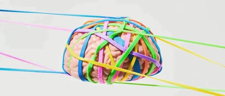
Brain wrapped in rubbberbands to demonstrate neuroplasticity

Does Brain Plasticity Increase After a Head Injury?

Medically reviewed by Nancy Hammond, M.D.
By James Roland for healthline.com on July 28, 2022

 
Brain plasticity, also called neuroplasticity, refers to the brain’s ability to adapt its structure and function in response to changes, such as a head injury or aging. Brain plasticity also involves the formation of new connections between neurons (brain cells).

The brain’s ability to reorganize these features after an injury affects the nature of post-injury recovery.

The severity of the injury goes a long way toward determining how the brain responds. But it’s often possible to boost brain plasticity with interventions and rehabilitation during the healing process.

What is brain plasticity?
Brain plasticity is a term that refers to the brain’s ability to restructure and reconfigure itself in response to change.

Change that can influence the brain comes in several forms. Expected changes include learning, experience, and aging. Unexpected changes include things like stroke and head injury.

Neuroplasticity has long been observed in children. It involves a process called neurogenesis, which is the formation of new neurons in the brain (and elsewhere in the nervous system).

There are two basic types of brain plasticity: structural and functional.

Structural Plasticity
Structural plasticity refers to the way the brain’s physical structure changes in response to learning.

For example, a small 2018 studyTrusted Source showed that healthy adults who participated in balance training twice a week, for 12 weeks, experienced thickening in certain areas of the brain involved in spatial orientation.

A 2016 study examined neuroplasticity in people learning to read Braille. It found that over the course of daily lessons, for 3 weeks, study participants developed increased connectivity in regions of the brain involved in processing sensations like touch.

Functional Plasticity
Functional plasticity refers to the brain’s ability to heal itself after injury. To achieve this, healthy regions of the brain adapt to take over certain functions that the damaged parts of the brain used to perform. This makes functional plasticity especially relevant for people recovering from head injuries.

A 2017 review of studies examining the role of neuroplasticity in stroke recovery found that a stroke can actually trigger neuroplasticity in certain areas. Neuroplasticity plays a role as the brain tries to resume regular functions, like speaking and controlling the movement of limbs.
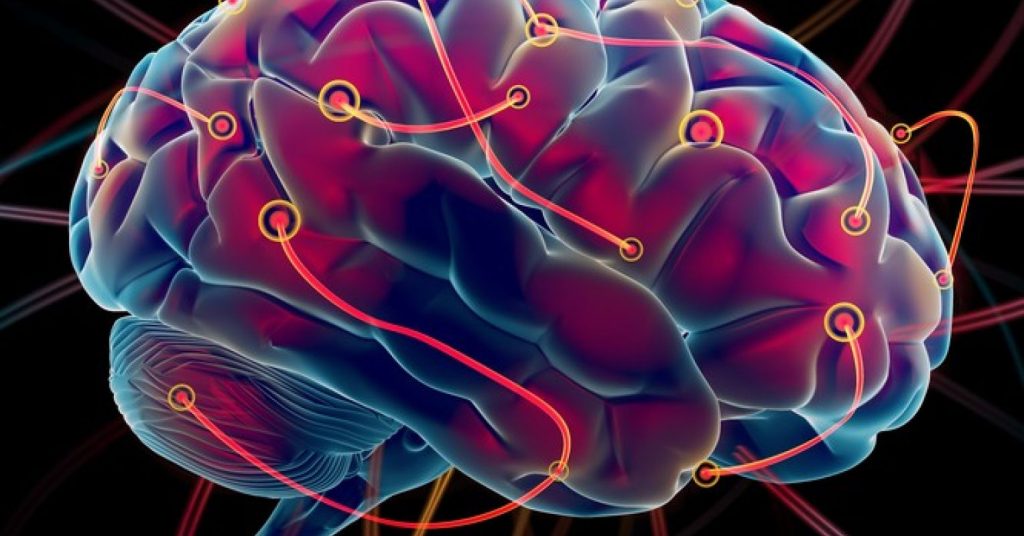
 
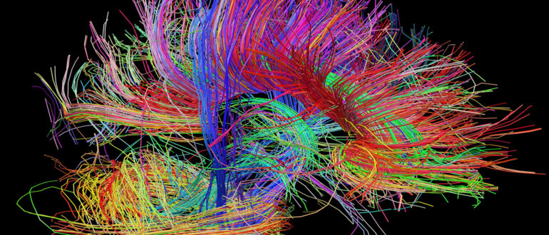
Brain image with new wiring

Can brain plasticity help you heal after a TBI?

 
A traumatic brain injury (TBI) refers to changes in brain function or brain health caused by an external force, such as a serious blow to the head.

The Centers for Disease Control and Prevention (CDC)Trusted Source reports that there were more than 220,000 TBI-related hospitalizations in 2019 and more than 64,000 TBI-related deaths the following year.

A TBI differs from a nontraumatic brain injury, also known as an acquired brain injury. Acquired brain injuries are those caused by internal factors, such as a stroke, which can damage brain tissue and affect muscle control, speech, cognition, and other functions.

When spontaneous brain plasticity doesn’t occur, it’s sometimes possible to boost neuroplasticity artificially.

 
A 2020 review of neuroplasticity therapies to treat stroke survivors suggests that approaches such as brain stimulation therapy and virtual reality might help enhance brain plasticity. It may also be possible to transfer nerves from healthy parts of the brain to injured parts.

Similarly, a 2017 review of studies on cognitive rehabilitation following TBI, suggests that memory and other thinking skills may be recovered to some degree with the help of cognitive rehabilitation. The studies showed how cognitive rehabilitation helped to modify damaged neural connections and various brain functions.

Does a brain injury increase neuroplasticity?

Because different regions of the brain are responsible for different functions, the location and severity of an injury determine which functions are affected and to what degree.

For example, certain areas of the brain are responsible for your ability to move certain parts of the body, like your left arm or your right foot.

This is where brain plasticity can help you heal after a brain injury. Just as exercise and learning can enhance brain structure and function, the body’s natural healing and recovery process after an injury can also increase neuroplasticity.

When neurons die due to injury, the brain naturally responds within a few days by developing new neural networks and recruiting various types of cells to take the place of those damaged or killed in the injury.

The extent to which neuroplasticity occurs depends on an individual’s age, the location of the injury, and other factors.

Does age matter after brain injury?

Whether it’s a brain injury or a broken wrist, being younger is always an advantage when it comes to recovery.

A 2008 studyTrusted Source of TBI survivors noted that disability scores following a TBI tended to be significantly better among younger TBI survivors compared with older individuals, even when those older survivors had less severe injuries. And the younger patients improved more in the first 5 years after the injury.

A 2019 report notes that because age affects neuroplasticity, the need for more strategies and therapies to compensate for age-related changes should be a higher priority in the face of an aging population.

Can you see brain plasticity on an MRI?

One of the most useful tools in diagnosing the impact of a TBI, stroke, or other injury or illness affecting the brain is magnetic resonance imaging (MRI).

An MRI can detect many changes in brain structure and function. Current technology is far from perfect, but it’s continuing to improve.

A 2021 articleTrusted Source suggests that advanced MRI techniques are helping doctors develop a more accurate picture of mild TBIs. This may help improve the treatment and understanding of mild TBIs in the future.

A newer type of MRI, called functional MRI (fMRI), can help doctors observe brain activity, not just brain structure. This may be particularly helpful in studying brain damage and recovery.

A 2017 studyTrusted Source of neuroimaging after TBI notes that fMRI can detect changes in thinking skills, emotions, and the course of neuroplasticity after an injury to the brain. The study says that fMRI is a helpful tool in assessing the damage caused by TBI and tracking brain changes during recovery.

But fMRI, the study says, will need to be accompanied by other data if it’s going to inform treatment decisions. This includes information gathered during cognitive-behavioral evaluations and other assessments.
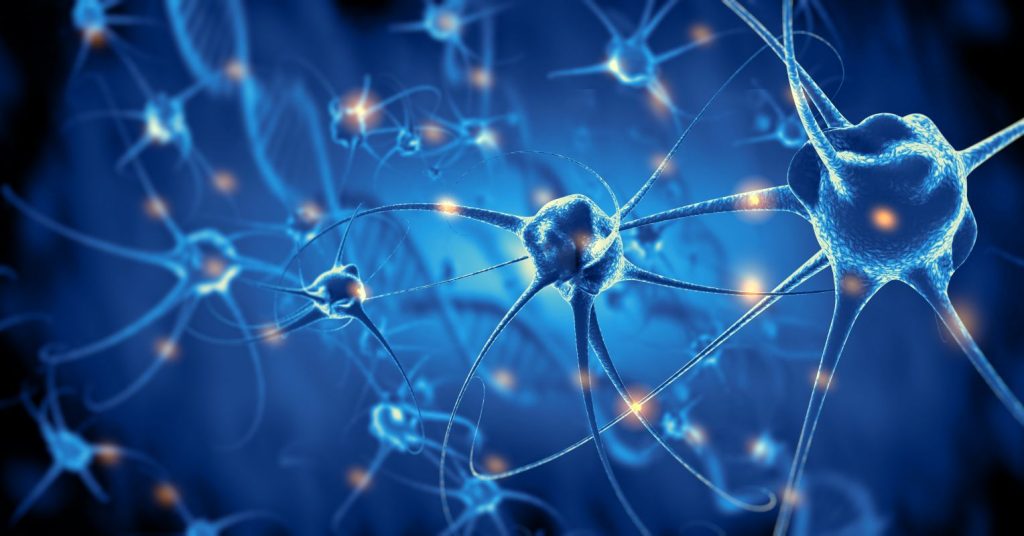
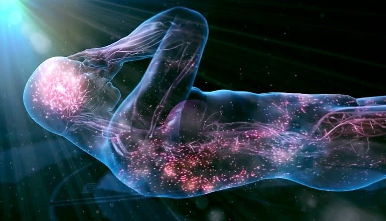
Image Neurons reconnecting

How long does it take to heal after a TBI?

 
The time necessary to heal from a TBI can vary considerably from one person to the next. This is based mostly on the seriousness of the injury, as well as its location, the age of the individual, and that person’s overall physical and mental health.

A full recovery from a mild TBI can be expected in about 3 months. People with a moderate TBI will take longer to heal and will typically need cognitive rehabilitation, physical therapy, and other interventions.

Predicting the degree and length of recovery from a severe TBI is very difficult, and should be done on a case-by-case basis.

Takeaway

Brain plasticity after a head injury is when brain functions thought to be lost due to damage begin to be adopted by other, healthy brain tissue.

While not all functions can be reorganized or reestablished completely, the brain’s remarkable adaptability can often help people who had a stroke, traumatic brain injury, or other harmful events recover some function.

Brain plasticity can be encouraged through cognitive therapy, physical therapy, and other treatments.

CLICK HERE to read the original article
 

Brain image (Photo/Courtesy of USC Stevens Institute for Neuroimaging and Informatics)

Brain image (Photo/Courtesy of USC Stevens Institute for Neuroimaging and Informatics)

Researchers Create Maps of the Brain After Traumatic Brain Injury

Anne Warde, UC Irvine, June 17, 2022

 
Scientists from the University of California, Irvine have discovered that an injury to one part of the brain changes the connections between nerve cells across the entire brain.

The new research was published this week in Nature Communications.

Every year in the United States, nearly two million Americans sustain a traumatic brain injury (TBI). Survivors can live with lifelong physical, cognitive and emotional disabilities. Currently, there are no treatments.

One of the biggest challenges for neuroscientists has been to fully understand how a TBI alters the cross-talk between different cells and brain regions.

In the new study, researchers improved upon a process called iDISCO, which uses solvents to make biological samples transparent. The process leaves behind a fully intact brain that can be illuminated with lasers and imaged in 3D with specialized microscopes.

With the enhanced brain clearing processes, the UCI team mapped neural connections throughout the entire brain. The researchers focused on connections to inhibitory neurons, because these neurons are extremely vulnerable to dying after a brain injury. The team first looked at the hippocampus, a brain region responsible for learning and memory.

Then, they investigated the prefrontal cortex, a brain region that works together with hippocampus. In both cases, the imaging showed that inhibitory neurons gain many more connections from neighboring nerve cells after TBI, but they become disconnected from the rest of the brain.

“We’ve known for a long time that the communication between different brain cells can change very dramatically after an injury,” said Robert Hunt, PhD, associate professor of anatomy and neurobiology and director of the Epilepsy Research Center at UCI School of Medicine whose lab conducted the study, “But, we haven’t been able to see what happens in the whole brain until now.”

To get a closer look at the damaged brain connections, Hunt and his team devised a technique for reversing the clearing procedure and probing the brain with traditional anatomical approaches.

The findings surprisingly showed that the long projections of distant nerve cells were still present in the damaged brain, but they no longer formed connections with inhibitory neurons.

“It looks like the entire brain is being carefully rewired to accommodate for the damage, regardless of whether there was direct injury to the region or not,” explained Alexa Tierno, a graduate student and co-first author of the study. “But different parts of the brain probably aren’t working together quite as well as they did before the injury.”

The researchers then wanted to determine if it was possible for inhibitory neurons to be reconnected with distant brain regions.

To find out, Hunt and his team transplanted new interneurons into the damaged hippocampus and mapped their connections, based on the team’s earlier research demonstrating interneuron transplantation can improve memory and stop seizures in mice with TBI.

The new neurons received appropriate connections from all over the brain. While this may mean it could be possible to entice the injured brain to repair these lost connections on its own, Hunt said learning how transplanted interneurons integrate into damaged brain circuits is essential for any future attempt to use these cells for brain repair.

One of the biggest challenges for neuroscientists has been to fully understand how a TBI alters the cross-talk between different cells and brain regions. Image is in the public domain One of the biggest challenges for neuroscientists has been to fully understand how a TBI alters the cross-talk between different cells and brain regions. Image is in the public domain

“Our study is a very important addition to our understanding of how inhibitory progenitors can one day be used therapeutically for the treatment of TBI, epilepsy or other brain disorders,” said Hunt.

“Some people have proposed interneuron transplantation might rejuvenate the brain by releasing unknown substances to boost innate regenerative capacity, but we’re finding the new neurons are really being hard wired into the brain.”

Hunt hopes to eventually develop cell therapy for people with TBI and epilepsy. The UCI team is now repeating the experiments using inhibitory neurons produced from human stem cells.

“This work takes us one step closer to a future cell-based therapy for people,” Hunt said, “Understanding the kinds of plasticity that exists after an injury will help us rebuild the injured brain with a very high degree of precision. However, it is very important that we proceed step wise toward this goal, and that takes time.”

Jan C. Frankowski, PhD; Shreya Pavani; Quincy Cao and David C. Lyon, PhD also contributed to this study.

CLICK HERE to read the original article
 

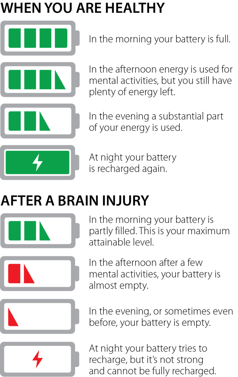
Fatigue After Brain Injury

By Katherine Dumsa, OTR/L, CBIS and Angela Spears, MA, CCC-SLP, DPNS, CBIS, Rainbow Rehabilitation Centers

 
Fatigue is a part of life that is experienced by everyone. Whether it is from a busy day at work, a demanding workout, or after paying attention to a long lecture, the term “I’m tired” is exceedingly common.

Fatigue and Traumatic Brain Injuries
For individuals with brain injuries, fatigue (sometimes referred to as cognitive fatigue, mental fatigue, or neurofatigue), is one of the most common and debilitating symptoms experienced during the recovery process. It can become a significant barrier to one’s ability to participate in the activities they want and need to do in daily life. It is reported that as many as 98% of people who have experienced a traumatic brain injury have some form of fatigue. Many report that fatigue is their most challenging symptom after brain injury. Reasons for the fatigue are not well understood but may include endocrine abnormalities, the need for the brain to work harder to compensate for brain injury deficits (in other words, inefficiency), or changes to brain structures.

Assessment Tools to Determine Fatigue Levels
Fatigue can be difficult to identify because it is not always reported by the patient or obvious to others. Clinicians use various self-report assessment tools to gain further information on a patient’s fatigue levels and the impact it has on their overall daily functioning. Two of the scales specifically designed for individual patients with brain injuries include the Barrow Neurological Institute Fatigue Scale (BNI) and the Cause of Fatigue Questionnaire (COF). Clinicians must also evaluate physical and mental changes, which can lead to depression and other psychiatric conditions following brain injury. The changes can commonly present as overwhelming fatigue.

Symptoms
Generally, those who have sustained brain injuries have described fatigue as a sense of mental or physical tiredness, exhaustion, lack of energy, and/or low vitality. Physical observations of fatigue include yawning, an appearance of confusion or “brain fog,” or easily losing attention and concentration. In more severe cases, it may present as forgetfulness, irritability, slurred speech, or dizziness. Emotions can become raw at this level of fatigue, affecting mood, motivation, and interaction with one’s social network. To manage fatigue effectively, individuals must learn to identify the symptoms of fatigue and how to modify activities that may trigger fatigue. Managing fatigue effectively will help decrease stress levels and improve overall performance for both work and home activities. Some fatigue-inducing activities include:

  • Working at a computer
  • Watching television excessively
  • Having a stimulating sensory environment
  • Concentrating on paperwork
  • Reading for long periods of time
  • Physically demanding tasks
  • Cognitively demanding tasks
  • Emotionally draining tasks
  •  
    Symptoms of fatigue can include:

  • Physical Symptoms: a pale or greyish pallor, glazed eyes, headaches, tension in muscles, shortness of breath, slower movement and speech, decreased coordination, or difficulty staying awake.
  • Cognitive Symptoms: increased forgetfulness, distractibility, decreased ability to follow directions, making an increased number of mistakes, decreased awareness of surroundings, or increased response time or lack of response.
  • Social/Emotional Symptoms: decreased ability to communicate effectively, decreased ability to engage in social activities, irritability, restlessness, emotional lability, increased negative thoughts, withdrawal, short answers, dull tone of voice, lack of motivation and interest, or difficulty engaging in activities of daily living.
  •  
    Fatigue Is Not Laziness
    In today’s multi-media society, we take in, absorb, and process large amounts of information every day. It can be difficult for family members or peers to understand the limitations caused by fatigue following a brain injury. Unfortunately, it can be mistaken for laziness or an unwillingness to participate in therapies and daily activities. It is important to understand that lacking the mental energy needed to complete tasks does not equate to lacking the desire to complete those tasks. Many individuals struggling with fatigue have motivation but lack the energy to keep up with daily demands.

    Coping Strategies Used to Ease Symptoms
    When managing fatigue, it is important to identify and treat physical factors that may be contributing to the fatigue. Recognizing early signs of fatigue and working with the patient so they understand how to respond to these is beneficial. By learning to recognize these triggers, one can learn coping strategies to successfully meet daily demands, ultimately increasing quality of life. These strategies include:

  • Having a Healthy Sleep Routine – This can be done by setting a sleep schedule of when to go to bed and when to wake, regardless of the day of the week. Establishing a strict routine using an alarm clock allows the brain proper rest. When rest is needed, aim for a “power nap” of 30 minutes maximum to avoid feeling over tired for the remainder of the day. Lack of sleep has a negative effect on our cognition, mood, energy levels, and appetite. The American Academy of Neurology reports that as many as 40% to 65% of people with mild traumatic brain injury complain of insomnia, so maintaining a sleep hygiene program is essential to recovery and to managing fatigue.
  • Practicing Energy Conservation – Pacing yourself each day, or prioritizing daily tasks to avoid becoming over-tired, can help with balancing out a busy schedule. Complete tasks that require the most mental effort earlier in the day with planned rest breaks in the afternoon or evening.
    Organizing daily activities – Utilize a checklist or planner to set a to-do list. Break up complex projects into manageable tasks. When completing these tasks, minimize environmental stimulation as much as possible.
  • Improving Health and Wellness – Increased overall health and wellness has been described as “energizing,” and research suggests that it can improve mood. Aim to exercise three to five times per week for a minimum of 30 minutes per session. Maintain a well-balanced diet rich in protein, fiber, and carbohydrates to help the brain and body stay fully energized.
  • Keeping a Fatigue Diary – This kind of diary can assist in monitoring changes and energy levels before and after daily activities. This tracking of fatigue can be used with your treatment team to help mitigate what may be increasing neurofatigue. Assessment and treatment of fatigue continues to be a challenge for clinicians and researchers. While there is no cure for fatigue, there are many ways to manage and overcome the symptom. Awareness and an open mind towards coping strategies will lessen the negative effects of fatigue and allow for meaningful participation in life.
     
    REFERENCES

  • Keough, A. 2016. Strategies to manage neuro-fatigue.
  • Cantor, J.H., Ashman, T., Gordon, W., Ginsberg, A., Engmann, C., Egan, M., Spielman, L., Dijkers, M., & Flanagan, S. (2008). Fatigue after traumatic brain injury and its impact on participation and quality of life. Journal of Head Trauma Rehabilitation. 23(1), 41-51.
  • Jang, S., & Kwon, H. (2016). Injury of the ascending reticular activating system in patients with fatigue and hypersomnia following mild traumatic brain injury. Medicine. 95(6). e2628.
  • Belmont, A., Agar, N., Hugeron, B. Gallais, C. & Azouvi, P., Fatigue and traumatic brain injury. Annales de Réadaptation et de Médecine Physique. 49(6). 370-374.
  • Johnson, G. (2000). Traumatic brain injury survival guide. Traverse City, MI.
  • Heins, J., Sevat, R., Werkhoven, C. (n.d.) Neurofatigue. Brain Injury Explanation.
  •  
    This article first appeared in the Summer 2018 issue of Rainbow Visions Magazine available at www.rainbowrehab.com.

    CLICK HERE to read the original article
     

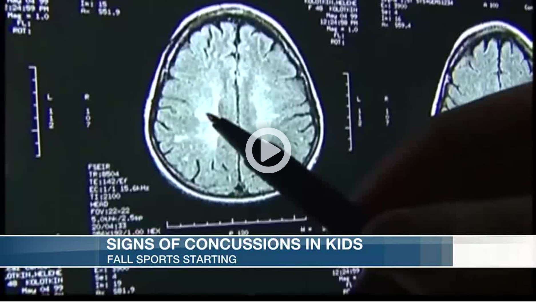
    Doctors Discuss Knowing the Signs of Concussion in Young Athletes

    By Adria Goins and Alex Onken, KSLA

     
    Thousands of students in the Arkansas/Louisiana/Texas (and across the nation) began fall sports over the last few weeks.

    However, with the new season here, comes a risk of injury. Football is the leading sport when it comes to concussions.

    The signs of a concussion are headache, fatigue and nausea. Parents are advised to then bring their child to a doctor right away if suspecting a possible concussion.

    “First diagnose it early and then after you diagnose it early make sure you avoid the triggers. So avoid extra screen time, over-exercising and just basically have 24 to 48 hours of cognitive physical rest,” said Dr. Kenneth Aguirre of Oschner-LSU Health Shreveport, who specializes in sports medicine.

    According to Dr. Charles Webb, also with Oschner-LSU Health and a sports medicine specialist, the topic of concussions and the potential risks of football comes up often.

    “I get that question a lot from parents. They want to know is it safe for my child to play high school or junior high, or even pee wee or popcorn football. and the question comes up because parents are worried about concussions. So my answer to them is if it were my child I wouldn’t let them play until they had an organized professional coach teaching them both how to hit and receive a hit.”

    Young athletes are usually taught how to hit and receive a hit around junior high. Dr. Webb said parents should put their children in club soccer or flag football in contrast to popcorn or pee wee football.

    “It’s much safer and you’re less likely to get hit in the head,” he said. “And you still get all the conditioning you need to play football later on in life.”

    In addition, doctors say keeping children awake when they have a concussion is a common misconception. Sleep is actually very good for the healing process.
     

    CLICK HERE to read the original article
     

    What Is a Neuropsych Evaluation?

    By Thomas A. Crosley, Crosley Law

     
    Brain injury can deeply impact how you think, make decisions, process information, and interact with others. When someone else caused your injuries, you deserve compensation for these losses.

    However, it can be hard to explain or document symptoms like memory loss, poor concentration, or impulsivity. In these cases, neuropsychological testing can help you, your medical team, and your personal injury lawyer understand the full effect of your traumatic brain injury (TBI).

    What Is Neuropsychology?

    The field of neuropsychology studies how our brains’ health impacts our emotions and behavior. Our brain is a remarkably complex organ, made up of nerves and tissues that help us feel, think, and perform everyday tasks. When there’s neurological dysfunction due to trauma, brain tumors, and diseases like Alzheimer’s, we may experience emotional and intellectual changes. Sometimes, these changes are subtle, like minor memory problems and mental “fogginess.” Other times, neurological issues create profound personality changes, cognitive deficits, and impaired decision-making.

    Different parts of the brain serve different functions. For example, the temporal lobe helps with our short-term memory, and the frontal lobe controls our memory, decision-making, and judgment. Depending on the location of your brain injury, you may exhibit different symptoms that affect your thinking, speech, vision, memory, and interpersonal relationships.

    A neuropsychological evaluation measures your emotional and cognitive abilities and compares them to the average person of your age, education, and background. An evaluation will typically consider a wide variety of factors, including:

  • Cognitive and intellectual abilities
  • Short-term and long-term memory
  • Executive functioning (your ability to make decisions and interpret information)
  • Speed of processing
  • Concentration and attention
  • Persistence and pace (your ability to finish tasks)
  • Gross and fine motor skills
  • Visual-spatial skills
  • Emotional functioning
  •  
    The evaluator may also look for other possible diagnoses, including depression, anxiety, or post-traumatic stress disorder. Finally, the evaluator will typically assess your performance and symptom validity; this process makes a neuropsych evaluation more objective than some other cognitive and mental assessments.

    Using Neuropsychological Testing to Assess the Impact of Brain Injury: A Case Study

    A neuropsychological report can help your doctors, lawyers, and mental health providers understand the full impact of your brain injury, which allows them to build effective treatment plans and fight to get you the compensation you deserve.

    Let’s look at a real-world example of how a neuropsych examination can help a TBI survivor’s legal claims. I represented a young man with an autism spectrum disorder who was struck by a delivery driver’s car while walking home from his job at a grocery store. During the collision, my client suffered significant brain injury, but the insurance company argued that his cognitive and memory deficits were due to his preexisting autism.

    To fight back, we consulted with his medical providers and a neuropsychologist who helped document his trauma-related symptoms and limitations. After mediation, we settled my client’s TBI claim for a significant amount.

    What Should I Expect During a Neuropsych Evaluation?

    During a neuropsychological evaluation, a team of clinicians, including a trained neuropsychologist, will give you a series of tests that assess your thinking abilities, language skills, memory, mental processing, and other abilities. You can expect to do a variety of tasks, including:

  • Answering questions about your daily routine and symptoms
  • Demonstrating your skills at reading, writing, math, and problem-solving
  • Identifying images
  • Recalling information after a time
  • Drawing pictures
  • Solving puzzles
  •  
    Some tests will be oral, while some will be written, computer-based, or task-driven. The precise tests used during your neuropsychological evaluation will vary depending on your diagnosis and other factors.

    However, not all neuropsychologists focus on brain injuries. A specialist who mainly works with dementia patients or another population might offer as detailed and insightful analysis when evaluating a TBI. If you are selecting a neuropsychologist, make sure they understand and regularly work with people with brain injury.

    How Long Does Neuropsych Testing Take?

    A neuropsych evaluation will take up to eight hours to complete. Typically, you’ll be able to take breaks as needed. If you become too tired or overwhelmed, the evaluator may split the testing over several days.

    What Happens After I Complete My Evaluation?

    Once you’ve completed your testing, the neuropsychologist will review your results, medical records, and other information to create a comprehensive report that discusses your cognitive abilities and limitations. If the neuropsych evaluation was scheduled as part of your TBI care plan, the process will include treatment recommendations and referrals to specialists, like speech therapy and counseling services.

    However, if an insurance company requested your neuropsych evaluation, it might serve a different purpose. Sometimes, “independent medical examinations,” including neuropsychological testing, are used to deny or reduce the value of a TBI survivor’s legal claims.

    For example, the insurance company may argue that your performance validity or symptom validity scores suggest you’re exaggerating symptoms. Rather than recommending treatment that will help you overcome your traumatic brain injury, the report will minimize your symptoms and suggest that you’re malingering (pretending your problems are worse than they are).

    To fight back, you’ll need to work with a personal injury lawyer who can carefully assess the evaluator’s methodology and identify issues and inconsistencies in their report. If you don’t already have an attorney, it’s a good idea to consult with a BIAA Preferred Attorney who has a documented track record of success.

    How Can I Prepare for Neuropsych Testing?

    While you can’t study for a neuropsych examination, there are some simple ways you can prepare for your appointment with the neuropsychologist:

  • Request an up-to-date list of your medications and prescriptions from your pharmacist or doctor
  • Get a good night’s sleep beforehand
  • Take your medications as prescribed
  • Eat a healthy meal before the exam
  • Dress comfortably for your day of testing
  • Wear your glasses or hearing aids, if needed
  •  
    Remember, as long as you are honest and give a good effort, you can’t “fail” a neuropsychological assessment.

    Worried About an Upcoming Neuropsych Evaluation? Consult with a BIAA Preferred Attorney

    If the insurance company schedules a neuropsych evaluation, it’s a good idea to consult with an experienced TBI lawyer. When you work with a BIAA Preferred Attorney, they can help you prepare for your examination, identify issues that may impact your legal claims, and fight back against an insurance company’s negative neuropsychological report.

    To find a TBI lawyer in your community, visit the BIAA Preferred Attorney page and click on “Narrow Your Search.” You’ll be able to filter Preferred Attorneys by their location and practice area.
     

    CLICK HERE to read the original article


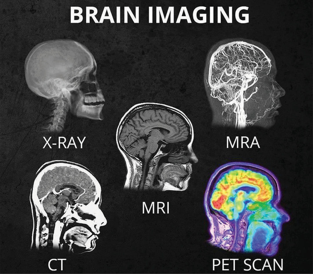
    What’s the difference between all the different head scans (X-Ray, CT, MRI, MRA, PET scan)? And what do they show in the head?

    Michael S. Tehrani, M.D.Follow Founder & CEO at MedWell Medical

     
    Ever wonder what’s the difference between all the different head scans (xray, CT, MRI, MRA, PET scan) and what they show in the head. Well wonder no more. The Dr. T easy to understand version…

    X-Ray: shows bone/skull only. Does not show the brain. Best used to detect if there are bone fractures.

    CT: a quick test. Shows brain but detail not great. Shows if any larger bleed, stroke, lesions, or masses.

    MRI: a long test. Shows brain and detail is great. Shows smaller bleeds, stroke, lesions, or masses.

    MRA:
    shows the flow of blood in the vasculature system of the brain. If there is vessel narrowing or blockage this test would show it.

    PET scan: shows how active different parts of the brain is. An active brain uses sugar as energy and pet scan detects how much sugar is being used by lighting up and turning different colors. The more sugar being used the more that area will light up and be different in colors. Cancer cells use the most sugar so cancer cells light up the most. PET scan is used to see if there are cancer cells. (Cancer cells replicate at a very fast and uncontrolled rate hence use a lot of sugar to allow that replication hence why they light up so much).

    CLICK HERE to download the original article
     

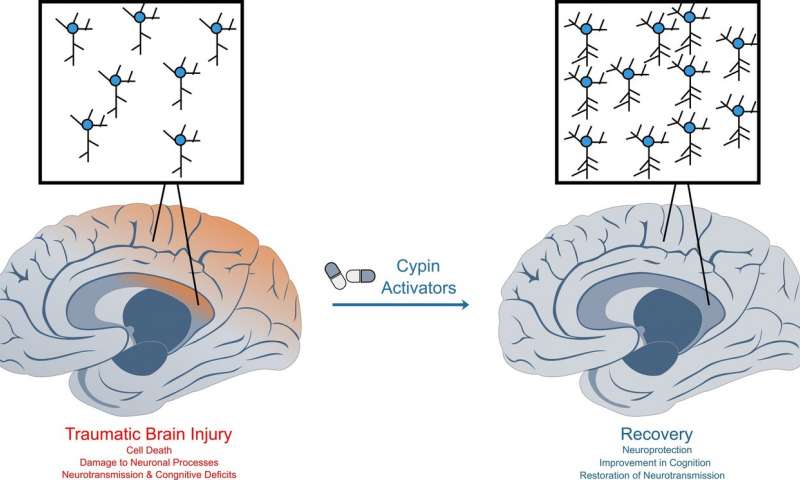
    Traumatic brain injury causes widespread damage to neurons, leading to deficits in learning and memory. Cypin activators restore neuronal survival and function in mice, allowing for normal learning and memory. Credit: Mihir Patel/Rutgers University-New Brunswick

    Traumatic brain injury: Discovery of two molecules could lead to new drug treatments

    By Todd B. Bates, July 27, 2018, Rutgers University

    After 10 years of research, a Rutgers-led team of scientists has identified two molecules that protect nerve cells after a traumatic brain injury and could lead to new drug treatments.

    The molecules promote full recovery after traumatic brain injury (TBI) in mice, according to the study published online in Neurobiology of Disease. Traumatic brain injury is the leading cause of death for people under 45 years old in the United States and is associated with disability, early-onset dementia, cognitive disorders, mental illness and epilepsy.

    Nearly all approaches for treating TBI focus on trying to prevent neurons, or nerve cells, from degenerating or on attempting to promote their survival, the study notes. TBI typically alters neural circuits within injured brain regions.

    “The big issue with treatment after TBI is that there are no drugs that work well on patients to restore memory, and we’re targeting reconnectivity of neural circuitry,” said Bonnie L. Firestein, senior author of the study and a professor in the Department of Cell Biology and Neuroscience at Rutgers University-New Brunswick. “That means we want our neurons to function properly and connect with other neurons. We want to allow people to retain their cognition and ability to remember and learn, so our angle is novel.”

    The researchers studied the protein cypin, an enzyme that breaks down guanine, which is an important building block for DNA and RNA in cells. The scientists previously showed that cypin is involved in promoting the proper shape in neurons and “keeping them happy,” Firestein said. This study found that speeding the breakdown of guanine protects neurons from injury and retains brain functioning.

    Scientists at Rutgers-New Brunswick, University of Pennsylvania, Fox Chase Chemical Diversity Center Inc. and Columbia University want to develop drugs from the molecules for further studies.

    Read the original article
     

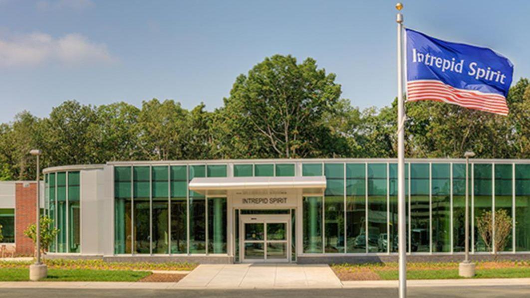
    The Intrepid Spirit traumatic brain injury treatment center is slated to open April 2 at Camp Pendleton. (Courtesy Naval Hospital Camp Pendleton) (Photo/iStock)

    Brain injury center to open at Marine base

    By Linda McIntosh, March 27, 2018, sandiegouniontribune.com

    A brain injury treatment center for military personnel will open its doors April 2 near the Naval Hospital Camp Pendleton.

    The $11.5 million Intrepid Spirit center is the seventh of nine such facilities at military bases across the country. It is funded by the New York-based nonprofit Intrepid Fallen Heroes Fund founded in 2000 by Zachary Fisher, who also started the Fisher House Foundation for military families.

    The center will operate as a part of Naval Hospital Camp Pendleton to treat active-duty military patients who suffer from the physical and psychological effects of brain injury. The center will also provide education and other resources on brain injury for veterans and the wider community.

    The center will expand the hospital’s existing program at the Concussion Care Clinic, which has served more than 2,000 patients since 2014. An estimated 550-600 new patients are expected to be referred to the center each year.

    “The facility will offer interdisciplinary, state-of-the-art evaluation of service members using clinical, laboratory and imaging resources to guide treatment,” said Cmdr. Paul Sargent, medical director of the Intrepid Spirit center, Naval Hospital Camp Pendleton.

    The center’s specialty rehabilitation and therapy programs will focus on providing service members strategies to improve recovery from physical, emotional and spiritual injuries.

    “We know that being able to be close to home, surrounded by loved ones, is a crucial part of the recovery process, so we are opening centers on the West Coast this spring at Camp Pendleton and also at Joint Base Lewis-McChord in Washington in order that service members who need treatment do not have to uproot themselves and their families to get it,” said David Winters, president of the Intrepid Fallen Heroes Fund.

    Two teams of clinicians will serve the clinic. Their specialties range from neurology, physical medicine and rehabilitation, psychiatry, trauma psychology, neuropsychology and pain psychology to physical and occupational therapy, creative arts therapy and neuro-optometry.

    “Our approach is a broadly collaborative center for preventing, treating and researching head trauma and injury to the brain,” Sargent said.

    The Intrepid Spirit center includes research, education and clinical staff from the Defense and Veterans Brain Injury Center, which is part of the Department of Defense’s Health Agency.

    “Teaching Marines, sailors and their commands about the risks of head injury, how to mitigate concussions and how to understand Traumatic Brain Injury signs and symptoms, along with how to improve readiness is a major goal of our TBI training,” said Regional Education Coordinator Clint Pearman, a certified brain injury specialist with the Defense and Veterans Brain Injury Center.

    Pearman provides outreach, education, training and resources for medical personnel, military commands, service members, veterans and family members and civilian community groups from the Camp Pendleton area up to northern California.

    The center’s design is based on the original National Intrepid Center of Excellence, which opened in 2010 at the Walter Reed National Military Medical Center in Bethesda, Md., operated by the Department of Defense.

    “There are hundreds of thousands of U.S. service members who continue to suffer from traumatic brian injury and other psychological health conditions,” Winters said. “The Intrepid Fallen Heroes Fund has tried to help these brave men and women get the best care available, so we made it our mission to build nine Intrepid Spirit centers that provide comprehensive, state-of-the-art treatment.”

    The clinic’s ground breaking was last May and a grand opening ceremony will be held at 11 a.m. April 4 at the Intrepid Spirit Center.

    For information about base access, visit pendleton.marines.mil/About/Base-Information/Base-Access.

    Read the original article


    By Alyssa Navarro, Tech Times (August 23, 2016) — Federal health regulators in the United States approved on Monday the use of two new computer softwares as cognitive screening tests for traumatic head injury patients.

    Known as ImPACT or the Immediate Post-Concussion Assessment and Cognitive Testing (ImPACT), the new testing device, as well as a similar test designed for children, can be used by doctors to evaluate signs and symptoms of head injuries that could indicate concussion.

    ImPACT is designed for patients aged 12 to 59 years old, while ImPACT Pediatric is intended for children aged 5 to 11 years old, officials said. Licensed health care professionals are the only ones allowed to perform the analysis and interpret the results.

    The software can be accessed easily because it runs on both desktop computers and laptops, according to the U.S. Food and Drug Administration (FDA). Both tests the first ever devices permitted by the FDA to assess cognitive function after experiencing a possible concussion. They are designed to be part of medical evaluations in hospitals.

    Although ImPACT and ImPACT Pediatric will definitely be useful for doctors, both tests are not meant to diagnose concussions or determine treatments that are appropriate for such cases, the FDA said.

    Instead, both devices are only designed to test cognitive skills such as reaction time, memory and word recognition. All of these can be impacted by head injuries. Afterwards, the results are compared to a patient’s pre-injury baseline scores or an age-matched control database, the FDA said.

    Dr. Carlos Peña, director of the neurological and physical medicine division at the Center for Devices and Radiological Health, acknowledges that the two testing devices can provide useful information that can aid doctors in the evaluation of people who are experiencing potential signs of concussion.

    However, Peña says that clinicians should not completely depend on the tests alone to rule out concussion or to decide whether a player with a head injury should return to a game.

    Statistics from the Centers for Disease Control and Prevention (CDC) reveal that traumatic brain injuries are responsible for more than 2 million visits to the emergency room in the country annually. Traumatic brain injuries also account for more than 50,000 deaths in America every year.

    Cases of head injury among kids have been increasing. In May, a CDC report showed that from January 2001 to December 2013, approximately 214,883 children aged 14 years old and below were brought to emergency departments due to head injuries.

    Read the original article
     

    UCSF Researchers Advocate Prioritizing Teens for Education and Prevention

    by Scott Maier (August 17, 2016) — The number of Americans diagnosed with concussions is growing, most significantly in adolescents, according to researchers at UC San Francisco. They recommend that adolescents be prioritized for ongoing work in concussion education, diagnosis, treatment and prevention.

    The findings appear online August 16, 2016, in the Orthopaedic Journal of Sports Medicine.

    “Our study evaluated a large cross-section of the U.S. population,” said lead author Alan Zhang, MD, UCSF Health orthopaedic surgeon. “We were surprised to see that the increase in concussion cases over the past few years mainly were from adolescent patients aged 10 to 19.”

    Concussions are a form of mild traumatic brain injury resulting in transient functional and biochemical changes in the brain. They can lead to time lost from sports, work and school, as well as significant medical costs.

    Though symptoms resolve in most concussion patients within weeks, some patients’ symptoms last for months, including depression, headache, dizziness and fogginess. Neuroimaging and neuropathological studies also suggest there may be chronic structural abnormalities in the brain following multiple concussions.

    Recent studies have shown an increase in traumatic brain injuries diagnosed in many U.S. emergency departments. Smaller cohort studies of pediatric and high school athletes also have indicated a rise in concussions for certain sports, such as football and girls’ soccer. However, this is the first study to assess trends in concussion diagnoses across the general U.S. population in various age groups.

    In this study, Zhang and his colleagues evaluated the health records of 8,828,248 members of Humana Inc., a large private payer insurance group. Patients under age 65 who were diagnosed with a concussion between 2007-2014 were categorized by year of diagnosis, age group, sex, concussion classification, and health care setting of diagnosis (emergency department or physician’s office).

    Overall, 43,884 patients were diagnosed with a concussion, with 55 percent being male. The highest incidence was in the 15-19 age group at 16.5 concussions per 1,000 patients, followed by ages 10-14 at 10.5, 20-24 at 5.2 and 5-9 at 3.5.

    The study found that 56 percent of concussions were diagnosed in the emergency department, 29 percent in a physician’s office, and the remainder in urgent care or inpatient settings. As such, outpatient clinicians should have the same confidence and competence to manage concussion cases as emergency physicians, Zhang said.

    A 60 percent increase in concussions occurred from 2007 to 2014 (3,529 to 8,217), with the largest growth in ages 10-14 at 143 percent and 15-19 at 87 percent. Based on classification, 29 percent of concussions were associated with some loss of consciousness.

    A possible explanation for the significant number of adolescent concussions is increased participation in sports, said Zhang, MD, who is also assistant professor of orthopaedic surgery at UCSF. It also may be reflective of an improved awareness for the injury by patients, parents, coaches, sports medical staff and treating physicians.

    For example, the U.S. Centers for Disease Control and Prevention “HEADS UP” initiative has caused numerous states such as California to alter guidelines for youth concussion treatment.

    Many medical centers also are establishing specialty clinics to address this, which could be contributing to the increased awareness. At UCSF, the Sports Concussion Program evaluates and treats athletes who have suffered a sports-related concussion. The team includes experts from sports medicine, physical medicine and rehabilitation, neuropsychology and neurology. Their combined expertise allows for evaluation, diagnosis and management of athletes with sports concussions, helping them safely recover and return to sports.

    Other UCSF orthopaedic surgery contributors to the Orthopaedic Journal of Sports Medicine study were senior author Carlin Senter, MD, associate professor; Brian Feeley, MD, associate professor; Caitlin Rugg, MD, resident; and David Sing, clinical research associate.

    UC San Francisco (UCSF) is a leading university dedicated to promoting health worldwide through advanced biomedical research, graduate-level education in the life sciences and health professions, and excellence in patient care. It includes top-ranked graduate schools of dentistry, medicine, nursing and pharmacy; a graduate division with nationally renowned programs in basic, biomedical, translational and population sciences; and a preeminent biomedical research enterprise. It also includes UCSF Health, which comprises two top-ranked hospitals, UCSF Medical Center and UCSF Benioff Children’s Hospital San Francisco, and other partner and affiliated hospitals and healthcare providers throughout the Bay Area.

    Read the original article
     

    Serving the Brain Injury Community Since 1983