News


New Rules to Protect Your Kid’s Noggin

May 25, 2019, Parents Magazine

 
Children bonk their head all the time when they’re wrestling with siblings, playing soccer, and just being clumsy-and it’s easy to worry that a bump could turn into something bigger. After all, more than 800,000 kids in the U.S. get a concussion every year. For the first time, the Centers for Disease Control and Prevention has released specific “return to learn” and “return to play” guidelines for head injuries, based on 25 years of research. One doctor shares the big takeaways.

ALWAYS take any injury beyond a light head bump seiously. A concussion occurs when a bump, blow, or jolt to the head or a hit to the body makes the brain bounce or twist in the skull. This creates chemical changes and can sometimes damage brain cells. “If your child complains of a headache or dizziness, is nauseous or vomiting, appears dazed, or sleeps more or less than usual, it’s time to get a doctor’s evaluation,” says Dennis Cardone, D.O., associate professor of orthopedic surgery and pediatrics and co-director of the NYU Langone Concussion Center. Even toddlers can get a concussion from a tumble, so look for changes in their behavior such as not wanting to nurse or eat or losing interest in toys.

If diagnosed with a concussion, your child will need menlal rest, says Dr. Cardone. That means taking a break from all activities for two to three days, and after that, starting with light aerobic activity. He may need to attend school for only half the day or do little to no homework (he won’t mind this rule!). However, he shouldn’t return to any sports or strenuous activities that have a high risk of falling or contact (think: field hockey, gymnastics, climbing a tree) until he’s been cleared by his doctor, which should be within a few weeks.

Download the original article PDF


These brain facts dispel many brain myths based on outdated knowledge. Learn how the brain works, for better (or worse). The original article, link at the bottom of the page, has the fact citations


72 Amazing Human Brain Facts (Based on the Latest Science)

Created by Deane Alban | Reviewed by Patrick Alban, DC
Last updated on February 6, 2019
BeBrainFit.com

 
There are a lot of myths and misinformation about the brain that pass as brain “facts.”

This is somewhat understandable: The study of the human brain is one of the least explored areas in science and even experts agree that there is more we don’t know about the brain than we currently do know.

In recent years, our knowledge of the brain has exploded — most of what we know about the brain has been discovered in just the last 15 years.

So the real brain facts haven’t always entered mainstream awareness yet.

This is a newly expanded and updated article.

We will continue to update this as new information comes to light, but for right now, here are 72 fascinating brain facts, all backed by the latest science.

    HUMAN BRAIN FACTS BY THE NUMBERS

  1. The typical brain comprises about 2% of the body’s total weight, but uses 20% of its total energy and oxygen intake.
  2. Your brain is 73% water. It takes only 2% dehydration to affect your attention, memory and other cognitive skills.
  3. Ninety minutes of sweating can temporarily shrink the brain as much as one year of aging does.
  4. Your brain weighs about three pounds. Sixty percent of the dry weight is fat, making the brain the most fatty organ in the body.
  5. Twenty-five percent of the body’s cholesterol resides within the brain. Cholesterol is an integral part of every brain cell. Without adequate cholesterol, brain cells die.
  6. No one knows for sure, but the latest estimate is that our brains contain roughly 86 billion brain cells.
  7. Each neuron can transmit 1,000 nerve impulses per second and make as many as tens of thousands of synaptic contacts with other neurons.
  8. A piece of brain tissue the size of a grain of sand contains 100,000 neurons and 1 billion synapses, all communicating with each other.
  9. All brain cells are not alike. There are as many as 10,000 specific types of neurons in the brain.
  10. Your brain needs a constant supply of oxygen. As little as five minutes without oxygen can cause some brain cells to die, leading to severe brain damage.
  11. Babies have big heads to hold rapidly growing brains. A 2-year-old’s brain is 80% of adult size.
  12. As any parent can attest, teenage brains are not fully formed. It isn’t until about the age of 25 that the human brain reaches full maturity.
  13. Brain information travels up to an impressive 268 miles per hour. This is faster than Formula 1 race cars which top out at 240 mph.
  14. Your brain generates about 12-25 watts of electricity. This is enough to power a low-wattage LED light.
  15. There’s a reason the brain has been called a “random thought generator.” The average brain is believed to generate up to 50,000 thoughts per day.
  16. Every minute, 750-1,000 milliliters of blood flows through the brain. This is enough to fill a bottle of wine or liter bottle of soda.
  17. Your brain can process an image that your eyes have seen for as little as 13 milliseconds — less time than it takes for you to blink.
  18.  
    FUN FACTS ABOUT BRAIN SIZE

  19. In general, men’s brains are 10% bigger than women’s, even after taking into account larger body size. However, the hippocampus, the part of the brain most strongly linked with memory, is typically larger in women.
  20. Albert Einstein’s brain weighed 2.71 pounds (1,230 grams) — 10% smaller than the average of 3 pounds (1,400 grams). However, the neuron density of his brain was greater than average.
  21. Neanderthal brains were 10% larger than our Homo sapiens brains.
  22. While humans have the largest brains proportional to body weight of all animals, we don’t have the biggest brains. That distinction belongs to sperm whales with 17-pound brains.
  23. Human brains have gotten significantly smaller over the past 10-20,000 years. The lost volume is equivalent to the size of a tennis ball.
  24. The hippocampus, the part of the brain considered the “memory center,” is significantly larger in London cab drivers. This is due to the mental workout they get while navigating the 25,000 streets of London.
  25.  
    THE EFFECTS OF THE MODERN LIFESTYLE ON THE BRAIN

  26. Chronic stress and depression are rampant in modern life. Either can cause measurable brain shrinkage.
  27. The modern diet is low in omega-3 essential fatty acids. Low levels of omega-3s result in brain shrinkage equivalent to two years of structural brain aging.
  28. Since the Victorian era, average IQs have gone down 1.6 points per decade for a total of 13.35 points.
  29. Technology has forced most of us to be prodigious multitaskers. But your brain can’t learn or concentrate on two things at once. What it can do is quickly toggle back and forth between tasks. But doing so decreases your attention span, ability to learn, short-term memory, and overall mental performance.
  30. Unexpectedly, millennials (aged 18 to 34) are more forgetful than baby boomers. They are more likely to forget what day it is or where they put their keys than their parents!
  31. Attention spans are getting shorter. In 2000, the average attention span was 12 seconds. Now, it’s 8 seconds. That’s shorter than the 9-second attention span of the average goldfish.
  32. Brain cells cannibalize themselves as a last ditch source of energy to ward off starvation. So, in very real ways, dieting, especially low-fat diets, can force your brain to eat itself.
  33. Over 140 proteins in the brain are negatively impacted by exposure to electromagnetic frequencies, the kind emitted by your cell phone and other electronic devices.
  34. Relying on GPS to navigate destroys your innate sense of direction, a skill that took our ancestors thousands of years to develop and hone. When areas of the brain involved in navigation are no longer used, those neural connections fade away via a process known as synaptic pruning.
  35.  
    BRAIN FACTS UPDATE: MYTHS DEBUNKED

  36. The popular myth that we use only 10% of our brains is flat-out wrong. Brain scans clearly show that we use most of our brain most of the time, even when we’re sleeping.
  37. There is no such thing as a left-brain or right-brain personality/skill type. We are not left-brained or right-brained; we are “whole-brained.”
  38. In spite of what you’ve been told, alcohol does not kill brain cells. What excessive alcohol consumption can do is damage the connective tissue at the end of neurons.
  39. The “Mozart effect” has been debunked. While listening to certain kinds of music can improve memory and concentration, there’s nothing unique about listening to Mozart.
  40. You may have heard that we have more brain cells than there are stars in the Milky Way, but this is not true. Best-guess estimates are that we have 86 billion neurons while there are 200-400 billion stars in the Milky Way.
  41. It’s often said that there are 10,000 miles of blood vessels in the brain when, actually, that number is closer to 400 miles. Still, a substantial amount!
  42. Contrary to the prevailing medical belief, having high total cholesterol is not bad for your brain. (See #5) In fact, high cholesterol actually reduces your risk of dementia.
  43. Until recently, it was a “fact” that you were born with a set level of intelligence and number of brain cells. But it has since been discovered that your brain has the capacity to change throughout your lifetime due to a property known as brain plasticity. The brain can continue to form new brain cells via a process known as neurogenesis.
  44.  
    FACTS ABOUT THE BRAIN AND MEMORY

  45. Memory is better thought of as an activity rather than being associated with a specific area of the brain. Any given memory is deconstructed and distributed in different parts of the brain. Then, for the memory to be recalled, it gets reconstructed from the individual fragments.
  46. Your brain starts slowing down at the ripe old age of 24, but peaks for different cognitive skills at different ages. In fact, at any given age, you’re likely getting better at some things and worse at others. An extreme case is vocabulary skills which may peak as late as the early 70s!
  47. If you were drinking alcohol and don’t remember what you did last night, it’s not because you forgot. While you are drunk, your brain is incapable of forming memories.
  48. It’s generally believed that people with exceptional memories are born that way, but this is rarely the case. Most memory masters will tell you that having an outstanding memory is a skill they developed by employing the best memory techniques.
  49.  
    FACTS ABOUT BRAIN FORM AND FUNCTION

  50. Human brain tissue is not dense. It’s very fragile — soft and squishy similar to the consistency of soft tofu or gelatin.
  51. The brain produces a half cup of fluid every day. It floats in this bath of cerebrospinal fluid which acts as a shock absorber to keep the brain from being crushed by its own weight.
  52. Sometimes half a brain is a good as a whole one. When surgeons operate to stop seizures, they remove or disable half of the brain in a procedure known as a hemispherectomy. Shockingly, patients experience no effect on personality or memory.
  53. Your brain has a pattern of connectivity as unique as your fingerprints.
  54. Although pain is processed in your brain, your brain has no pain receptors and feels no pain. This explains how brain surgery can be performed while the patient is awake with no pain or discomfort. Headache pain feels like it starts in your brain, but is caused by sensations from nearby skin, joints, sinuses, blood vessels or muscles.
  55. Brain freeze sure feels like pain in the brain but is an example of referred pain emanating from the roof of the mouth. Fortunately, brain freeze does not freeze brain cells because frozen brain cells rupture and turn to mush.
  56. The brains of introverts and extroverts are measurably different. MRIs reveal that the dopamine reward network is more active in the brains of extroverts while introverts’ brains have more gray matter.
  57. According to research done at Cambridge University, the order of letters in a word doesn’t matter much to your brain. As long as the first and last letters are in the right spot, your brain can rearrange the letters to form words as fast as you can read. This is why you can easily make sense out of this jumble of letters:
    Aoccdrnig to a rscheearch at Cmabrigde Uinervtisy, it deosn’t mttaer in waht oredr the ltteers in a wrod are, the olny iprmoetnt tihng is taht the frist and lsat ltteer be at the rghit pclae. The rset can be a toatl mses and you can sitll raed it wouthit porbelm. Tihs is bcuseae the huamn mnid deos not raed ervey lteter by istlef, but the wrod as a wlohe.
    Pretty amazing!
  58.  
    HOW THE BRAIN COMPARES TO A COMPUTER

  59. Your brain’s storage capacity is considered virtually unlimited. It doesn’t get “used up” like RAM in your computer.
  60. The latest research shows that the brain’s memory capacity is a quadrillion, or 1015, bytes. Astoundingly, this is about the same amount needed to store the entire internet!
  61. The human brain is capable of 1,016 processes per second, which makes it far more powerful than any existing computer.
  62. Researchers involved in the AI Impacts project have developed a way to compare supercomputers to brains — by measuring how fast a computer can move information around within its own system. By this standard, the human brain is 30 times more powerful than the IBM Sequoia, one of the world’s fastest supercomputers.
  63. Japan’s K computer is one of the most powerful computers in the world. When programmed to simulate human brain activity, it took 40 minutes to crunch the data equivalent to just one second of brain activity.
  64. &nbsp:
    EVIDENCE OUR BRAINS “COULD BE BETTER”

  65. There are almost 200 known cognitive biases and distortions that cause us to think and act irrationally.
  66. Memories are shockingly unreliable and change over time. Emotions, motivation, cues, context and frequency of use can all affect how accurately you remember something. This includes “flash bulb memories” which occur during traumatic events.
  67. Of the thousands of thoughts a person has every day, it’s estimated that 70% of this mental chatter is negative — self-critical, pessimistic, and fearful.
  68. Think you’re in control of your life? Don’t be so sure. Ninety-five percent of your decisions take place in your subconscious mind.
  69. A blood-brain barrier protects your brain by preventing many foreign substances in your vascular system from reaching the brain. But the barrier doesn’t work perfectly and many substances sneak through. Nicotine rushes into the brain in a mere 7 seconds. Alcohol, on the other hand, takes 6 minutes.
  70. Our brains crave mental stimulation, sometimes to a fault. Men especially would rather give themselves electric shocks than sit quietly in a room and think!
  71. Synesthesia is a condition where stimulation of one sense automatically evokes a perception of another sense. People with synesthesia might “taste” words, “smell” sounds, or see numbers as colors. While it’s not known exactly why this occurs, the prevailing theory is that these brains have hyper-connectivity between sensory areas in the brain.
  72. The human brain is extraordinarily complex and consequently can go awry in some spectacular ways. Some of the strangest disorders include exploding head syndrome disorder (hearing phantom explosions in your head), Capgras syndrome (thinking loved ones have been substituted by impostors, robots or aliens), and Cotard’s syndrome (believing you are dead).
  73. Savant syndrome is a condition where those with serious mental disabilities have an “island of genius.” The most common areas of genius fall into one of these categories: music, art, mathematics, mechanical, or spatial skills.
  74. Most savants are born that way, but a brain trauma can cause acquired savant syndrome where ordinary people suddenly develop genius-level abilities they didn’t have before.
  75. Brain cells need a constant supply of fuel to stay alive, yet they lack the ability to store energy. Fortunately, there’s a backup system. Your liver breaks down stored fat to produce ketone bodies that can be used as a substitute fuel when commonly-used blood glucose is not available.
  76.  
    BRAIN FACTS THAT ARE JUST PLAIN WEIRD

  77. The brain in your head isn’t your only brain. There’s a “second brain” in your intestines that contains 100 million neurons. Gut bacteria are responsible for making over 30 neurotransmitters including the “happy molecule” serotonin.
  78. Some scientists believe zombies could actually be created. They think it’s possible that a mutated virus or parasites could attack the brain and rapidly spread throughout large populations, essentially causing a “zombie apocalypse.”
  79. Users of Apple devices really are different than those who use Android products. (It’s not your imagination.) MRIs reveal that Apple products stimulate the “god spot” in their users’ brains — the same part of the brain activated by religious imagery in people of faith.
  80. Few facts about the brain are as strange as the posthumous story of Albert Einstein’s brain. The pathologist who performed Einstein’s autopsy kept the brain in a jar in his basement for 40 years. Eventually, he made a cross-country trip with the brain in a Tupperware container to deliver it to Einstein’s granddaughter.

Read the original article
 

Brainline Holiday Article Picture
 

15 Tips for Surviving — AND Enjoying — the Holidays with Brain Injury

By BrainLine

Flashing lights. Crowded stores. Loud family gatherings. The holiday season should be joyful, but it can often be overwhelming to someone who is living with brain injury.

If you are living with TBI, share these tips with your friends and family. If someone you love is living with TBI, the tips below can help you plan to make the holiday season happier and more relaxed for all of your friends and family.

These great ideas came from members of BrainLine’s wonderful online community.

  1. Identify — in advance, if possible — a quiet place to go at gatherings if you are feeling overwhelmed. This gives you a chance to take a break and lets your loved ones stay involved in the festivities.
  2. Avoid crowded stores and order gifts online instead.
  3. If you are shopping in stores, remember to make a list in advance and plan your trips on weekdays — either early in the morning or late at night when there are fewer crowds.
  4. Wear a cap with a brim or lightly tinted sunglasses to minimize the glare of bright lights in stores or flashing lights on a tree.
  5. Wear noise-reducing headphones or earbuds. These are also great gift ideas for loved ones with TBI if they don’t already have them.
  6. Ask a friend to go with you to stores or holiday parties. They can help you navigate crowds and anxiety-producing situations.
  7. Plan in advance as much as possible. And ask your hosts what their plans are so you aren’t surprised by anything.
  8. Volunteer to help with the holiday activities that you enjoy the most and are least stressful for you.
  9. Remember to ask for help and accept help if it is offered to you.
  10. Ask someone you trust to help you with a budget to avoid overspending on gifts.
  11. Take a nap if you need a break.
  12. Remember that it’s okay to skip the big parties and plan to celebrate in a way that makes you comfortable and happy.
  13. Check in advance to see if fireworks are part of outdoor celebrations — and skip them if they make you uncomfortable.
  14. If flashing lights bother you, ask your friends and family to turn off the flashing feature on Christmas tree lights or other decorations when you visit their homes.
  15. You can let your host know in advance that you may need to leave early. It will help you feel comfortable if you need to get home or to a quiet place and it can also help avoid any hurt feelings.

Read the original article
 

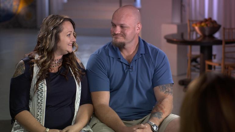
Libby and Tom Bates // CBS News

A brain disease best known for impacting football players who suffered concussions is now being found in soldiers

By Sharyn Alfonsi, September 16, 2018, CBS News

Until a few years ago, NFL players who struggled with severe depression, bouts of rage and memory loss in their retirement were often told they were just having a hard time adjusting to life away from the game. Doctors have since learned these changes can be symptoms of the degenerative brain disease CTE – chronic traumatic encephalopathy, caused by blows to the head.

As we first reported in January, CTE isn’t just affecting athletes, but also showing up in our nation’s heroes. Since 9/11 over 300,000 soldiers have returned home with brain injuries. Researchers fear the impact of CTE could cripple a generation of warriors.

When Joy Kieffer buried her 34-year old son this past summer, it was the end of a long goodbye.

Kieffer’s son, Sgt. Kevin Ash, enlisted in the Army Reserves at the age of 18. Over three deployments, he was exposed to 12 combat blasts, many of them roadside bombs. He returned home in 2012 a different man.

Joy Kieffer: His whole personality had changed. I thought it was exposure to all of the things that he had seen, and he had just become harder. You know, but he was — he was not happy.

Sharyn Alfonsi: So at this point, you’re thinking this decline, this change in my child is just that he’s been in war and he’s seen too much.

Joy Kieffer: Right.

Sharyn Alfonsi: Did he tell you about blasts that he experienced during that time?

Joy Kieffer: Uh-huh.

Sharyn Alfonsi: What did he–tell you?

Joy Kieffer: That they shook him. And he was having blackouts. And — it frightened him.

Ash withdrew from family and friends. He was angry. Depressed. Doctors prescribed therapy and medication, but his health began to decline quickly. By his 34th birthday, Sgt. Kevin Ash was unable to speak, walk or eat on his own.

Sharyn Alfonsi: Looking back on it now, was there anything you feel like he could’ve done?

Joy Kieffer: Uh-uh.

Sharyn Alfonsi: Because?

Joy Kieffer: Because it was– it– it was his brain. The thing I didn’t know was that his brain was continuing to die. I mean, before he went into the service he said, “you know, I could come back with no legs, or no arms, or even blind, or I could be shot, I could die,” but nobody ever said that he could lose his mind one day at a time.

His final wish was to serve his country one last time by donating his brain to science — a gesture he thought would bring better understanding to the invisible wounds of war.

Joy reached out to the VA-Boston University-Concussion Legacy Foundation Brain Bank where neuropathologist Dr. Ann McKee is leading the charge in researching head trauma and the degenerative brain disease CTE.

McKee has spent fourteen years looking at the postmortem brains of hundreds of athletes who suffered concussions while playing their sport.

Last summer, her findings shook the football world when she discovered CTE in the brains of 110 out of 111 deceased NFL players — raising serious concerns for those in the game today.

And when Dr. McKee autopsied Patriots tight-end Aaron Hernandez who killed himself after being convicted of murder, she found the most severe case of CTE ever, in someone under 30.

Now she’s seeing similar patterns in deceased veterans who experienced a different kind of head trauma — combat blasts. Of the 125 veterans’ brains Dr. Mckee’s examined, 74 had CTE.

Sharyn Alfonsi: I can understand a football player who keeps, you know, hitting his head, and having impact and concussions. But how is it that a combat veteran, who maybe just experienced a blast, has the same type of injury?

Dr. Ann McKee: This blast injury causes a tremendous sort of– ricochet or– or– a whiplash injury to the brain inside the skull and that’s what gives rise to the same changes that we see in football players, as in military veterans.

Blast trauma was first recognized back in World War I. Known as ‘shell shock,’ poorly protected soldiers often died immediately or went on to suffer physical and psychological symptoms. Today, sophisticated armor allows more soldiers to walk away from an explosion but exposure can still damage the brain — an injury that can worsen over time.

Dr. Ann McKee: It’s not a new injury. But what’s been really stumping us, I think, as– as physicians is it’s not easily detectable, right? It’s– you’ve got a lot of psychiatric symptoms– and you can’t see it very well on images of the brain and so it didn’t occur to us. And I think that’s been the gap, really, that this has been what everyone calls an invisible injury.

Dr. Ann McKee: This is the world’s largest CTE brain bank.

The only foolproof way to diagnose CTE is by testing a post-mortem brain.

Sharyn Alfonsi: So these are full of hundreds of brains…

Dr. Ann McKee: Hundreds of brains, thousands really…

Researchers carefully dissect sections of the brain where they look for changes in the folds of the frontal lobes – an area responsible for memory, judgement, emotions, impulse control and personality.

Dr. Ann McKee: Do you see there’s a tiny little hole there? That is an abnormality. And it’s a clear abnormality.

Sharyn Alfonsi: And what would that affect?

Dr. Ann McKee: Well, it’s part of the memory circuit. You can see that clear hole there that shouldn’t be there. It’s connecting the important memory regions of the brain with other regions. So that is a sign of CTE.

Thin slivers of the affected areas are then stained and viewed microscopically. It’s in these final stages where a diagnosis becomes clear as in the case of Sgt. Kevin Ash.

Sharyn Alfonsi: So this is Sergeant Ash’s brain?

Dr. Ann McKee: Right. This is– four sections of his brain. And what you can see is– these lesions. The, and those lesions are CTE And they’re in very characteristic parts of the brain. They’re at the bottom of the crevice. That’s a unique feature of CTE.

Sharyn Alfonsi: And in a healthy brain, you wouldn’t see any of those kind of brown spots?

Dr. Ann McKee: No, no, it would be completely clear. And then when you look microscopically, you can see that the tau, which is staining brown and is inside nerve cells is surrounding these little vessels.

Sharyn Alfonsi: And explain, what is the tau?

Dr. Ann McKee: So tau is a protein that’s normally in the nerve cell. It helps with structure and after trauma, it starts clumping up as a toxin inside the nerve cell. And over time, and even years, gradually that nerve cell dies.

Dr. Lee Goldstein has been building on Dr. McKee’s work with testing on mice.

Inside his Boston University lab, Dr. Goldstein built a 27-foot blast tube where a mouse – and in this demonstration, a model – is exposed to an explosion equivalent to the IEDs used in Iraq and Afghanistan.

Dr. Lee Goldstein: When it reaches about 25 this thing is going to go.

Dr. Goldstein’s model shows what’s going on inside the brain during a blast. The brightly colored waves illustrate stress on the soft tissues of the brain as it ricochets back and forth within the skull.

Dr. Lee Goldstein: What we see after these blast exposures, the animals actually look fine. Which is shocking to us. So they come out of what is a near lethal blast exposure, just like our military service men and women do. And they appear to be fine. But what we know is that that brain is not the same after that exposure as it was microseconds before. And if there is a subsequent exposure, that change will be accelerated. And ultimately, this triggers a neurodegenerative disease. And, in fact, we can see that really after even one of these exposures.

Sharyn Alfonsi: The Department of Defense estimates hundreds of thousands of soldiers have experienced a blast like this. What does that tell you?

Dr. Lee Goldstein: This is a disease and a problem that we’re going to be dealing with for decades. And it’s a huge public health problem. It’s a huge problem for the Veterans Administration. It’s a huge moral responsibility for all of us.

A responsibility owed to soldiers like 34-year-old Sgt. Tom Bates.

Sgt. Tom Bates: We were struck with a large IED. It was a total devastation strike.

Bates miraculously walked away from a mangled humvee — one of four IED blasts he survived during deployments in Iraq and Afghanistan.

Sharyn Alfonsi: Do you remember feeling the impact in your body?

Sgt. Tom Bates: Yes. Yeah.

Sharyn Alfonsi: What does that feel like?

Sgt. Tom Bates: Just basically like getting hit by a train.

Sharyn Alfonsi: And you were put back on the frontlines.

Sgt. Tom Bates: Yes.

Sharyn Alfonsi: And that was it?

Sgt. Tom Bates: Uh-huh

When Bates returned home in 2009, his wife Libby immediately saw a dramatic change.

Libby Bates: I thought, “Something is not absolutely right here. Something’s going on. For him to just lay there and to sob and be so sad. You know, what do you do for that? How do I– how do I help him? He would look at me and say, “If it wasn’t for you, I would end it all right now.” You know, I mean, like, what do you– what do you do– and what do you say to somebody who says that? You know I love this man so much. And —

Sharyn Alfonsi: You’re going to the VA, you’re getting help, but did you feel like you weren’t getting answers?

Sgt. Tom Bates: Yes.

Sharyn Alfonsi: And so you took it into your own hands and started researching?

Sgt. Tom Bates: I knew the way everything had gone and how quick a lot of my neurological issues had progressed that something was wrong. And I just– I wanted answers for it.

That led him to New York’s Mount Sinai Hospital where neurologist Dr. Sam Gandy is trying to move beyond diagnosing CTE only in the dead by using scans that test for the disease in the living.

Dr. Sam Gandy: By having this during life, this now gives us for the first time the possibility of estimating the true prevalence of the disease. It’s important to estimate prevalence so that people can have some sense of what the risk is.

In the past year, 50 veterans and athletes have been tested for the disease here. Tom Bates asked to be a part of it.

That radioactive tracer – known as t807 – clings to those dead clusters of protein known as tau, which are typical markers of the disease.

Through the course of a 20 minute PET scan, high resolution images are taken of the brain and then combined with MRI results to get a 360 degree picture of whether there are potential signs of CTE.

Scan results confirmed what Tom and Libby had long suspected.

On the right, we see a normal brain scan with no signs of CTE next to Tom’s brain where tau deposits, possible markers of CTE, are bright orange.

Dr. Sam Gandy: Here these could be responsible for some of the anxiety and depression he’s suffered and we’re concerned it will progress.

Sgt. Tom Bates: My hope is that this study becomes more prominent, and gets to more veterans, and stuff like that so we can actually get, like, a reflection of what population might actually have this.

There is no cure for CTE.

Dr. Gandy hopes his trial will lead to drug therapies so he can offer some relief to patients like Tom.

Dr. Ann McKee believes some people may be at higher risk of getting the disease than others.

While examining NFL star Aaron Hernandez’s brain she identified a genetic bio-marker she believes may have predisposed him to CTE.

A discovery that could have far-reaching implications on the football field and battlefield.

Sharyn Alfonsi: Do you think you will ever be your old self again?

Sgt. Tom Bates: I don’t ever see me being my old self again. I think it’s just too far gone.

Sharyn Alfonsi: So what’s your hope then?

Sgt. Tom Bates: Just to not become worse than I am now.

Since our story first aired, over 100 veterans have contacted Dr. Gandy to enroll in ongoing trials to identify whether they are living with CTE. And more than 300 have reached out to Dr. Mckee about donating their brains to research.

Read the original article

 

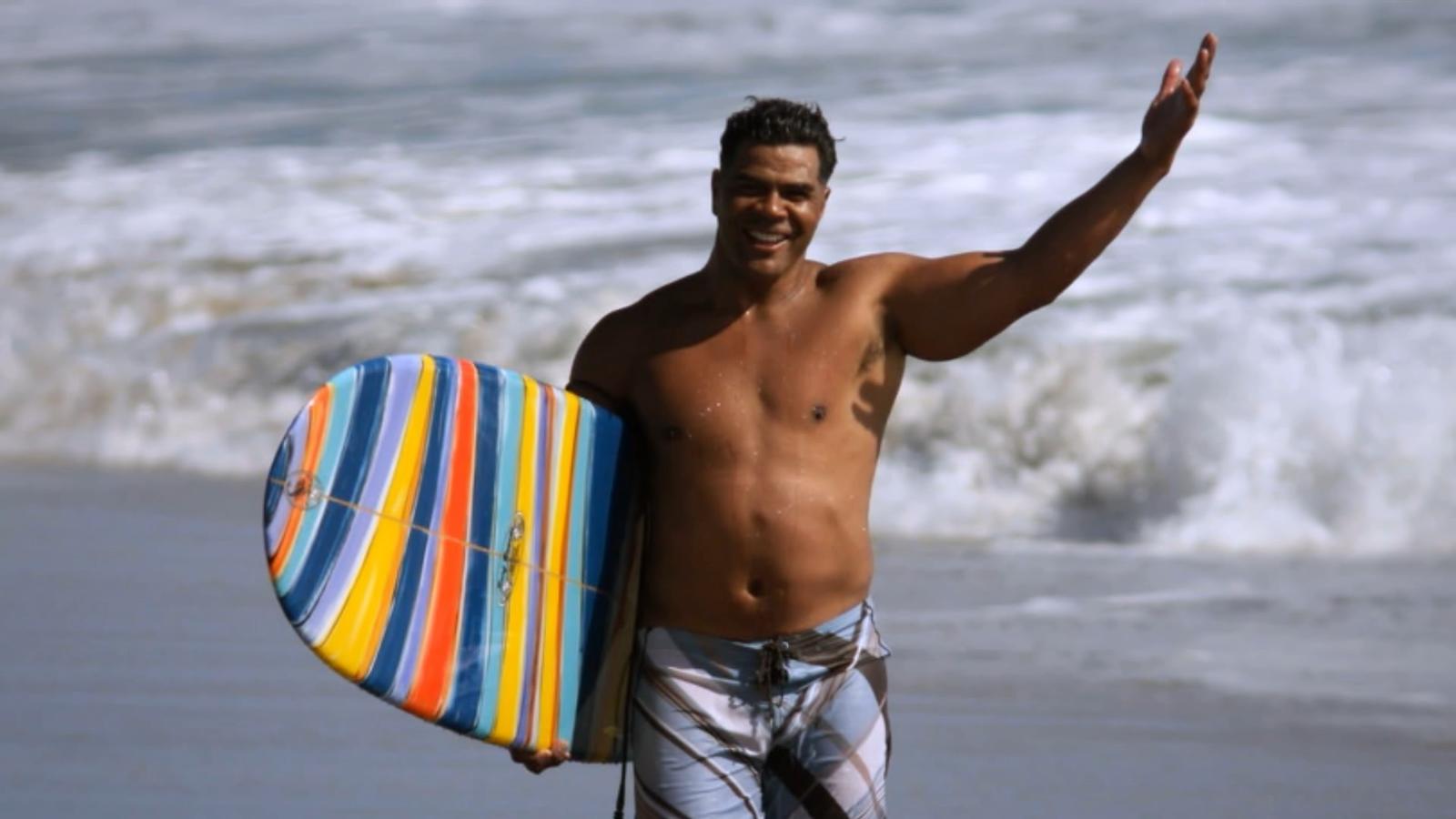
Junior Seau, shown at his beloved Pacific Ocean in the ESPN Films “30 for 30” documentary “Seau,” which premieres Thursday. (ESPN Films)

ESPN hits the mark with documentary ‘Seau’

By Tom Krasovic, September 20, 2018, San Diego Union Tribune

An aerial view of the Oceanside coast, in full sparkle and splendor below, grandly eases viewers into “Seau,” an ESPN Films documentary in the “30 for 30” series that debuts Thursday on the streaming service ESPN+.

It’s a sunny scene, the Pacific Ocean’s turquoise waves illuminated as they roll toward the white beach. The late Junior Seau told friends he found peace paddling on these waters, deep into his life alongside the town where he’d grown up.

Up at dawn with a yellow long-board and oar in hand, Seau had only a short walk from his beachfront home to the water.

Yet the former Chargers linebacker, role model and local philanthropist was then also writing in a journal of bouts with depression, memory loss and perceived guilt. There were headaches, too, and nights plagued by insomnia. “Buddy,” he’d told a friend and professional soccer player who’d suffered a brain injury from heading a ball, “I’ve had a concussion since I was 15.”

Diary entries also revealed feelings of humiliation and embarrassment over not living up to expectations of others and himself, and of feeling used by others.

“The world has nothing for me,” Seau pens in one entry, the cursive words all too legible.

One of Seau’s surviving adult children, after reading the grim line aloud, wonders why his father didn’t regard his family as something in this apparent world of nothing.

Why couldn’t they have been a lifeline for him to reach out and grasp?

“Seau,” produced and directed by Kirby Bradley, lets viewers draw their own conclusions about a complicated life that ended one May morning six years ago, at age 43, with a self-inflicted gunshot wound to the chest, but not before we hear from an array of family members, friends and experts in football and brain science.

At the end of the 90-minute film, themes of redemption and hope are raised.

“Let’s all walk from here being better for having known Junior Seau and the impact he had on our lives,” NFL quarterback Drew Brees, a former Chargers teammate of the Hall of Fame linebacker, concludes near the film’s end.

Former Chargers lineman Aaron Taylor notes that in death, Seau drew extraordinary attention to the link between head trauma and a degenerative brain disease, CTE, revealed in a tissue sample sent to a brain scientists at the family’s request.

Exciting beginnings and success are a thread to the film, followed often by bitter detours or hurtful endings.

Seau took to sports at Oceanside High with a passion that rivaled his stunning blend of size, speed and agility. If he was slamming into football ball-carriers or catching passes, scoring baskets or throwing the discus and shot, he was a “force of nature” for the green-and-white-clad Pirates, observers said.

A flood of football scholarship offers came to the small home where Seau and his brothers slept in a tiny garage.

Jubilation ensued when Seau chose USC, keeping him close to his parents and siblings and the tight-knit Samoan-American community in Oceanside. A similar celebration arose in 1990 when the Chargers drafted him fifth overall. “I’m a real momma’s boy,” Seau said, pulling on a blue team cap.

Playing for his beloved “Diego,” he led the long-struggling Chargers to the playoffs in just his third season, and their first Super Bowl two years later. “Now the world is gonna know the San Diego Chargers,” he told some 70,000 celebrants in Mission Valley after the team returned from claiming the 1994 AFC title in Pittsburgh.

The flip side?

If Oceanside lost a game in which he played, Junior lost his lunch money. It was the price his father exacted.

The thrill of signing with USC gave way to humiliation when a failed admittance test made him ineligible as a freshman. His father refused to talk to him in response, deeming the failure an embarrassment to the family. After a dominant junior year with USC, there would be no senior year. Making money was the next step, in no small part because he wanted to support his parents and other family members.

The Chargers couldn’t build upon their Super Bowl season, and the team’s constant losing wore on Seau.

When the Chargers traded him in the spring of 2003, after 13 seasons with the club, Seau was hurt that the team — Stay Unclassy, San Diego? — called not him but his agent to tell him the news. “I know that was hard on him,” said the agent, Steve Feldman.

Gina Seau was working for the Chargers in marketing when she first met Seau early in his NFL career.

She recalled “very kind eyes” and a “very soft voice” that almost “didn’t match the size and stature.”

The two would marry, but erratic behavior that Gina Seau linked to numerous football-related head injuries — “My head is on fire,” he told her — led to a divorce in 2002. The two remained friends. Believing that driving off a steep coastal cliff in October 2010 wasn’t an accident, Gina pleaded with her former husband to get help.

Here’s hoping that if there’s a “Seau II,” events yet to transpire bring more developments of redemption. Say, a cure for CTE.

Read the original article
 

Recently, I spoke to someone who may have accidentally taken a duplicate dose of insulin, resulting in a dangerously low blood sugar.
He knows that I am interested in assistive memory technology because I work with many patients with memory challenges. . So he asked if I could recommend a way to quickly record his insulin doses using his iPhone in order to prevent accidental overdosing.

I did some research on the many available apps for diabetes management. While I found many highly-rated apps, I tried to think of the simplest and easiest methods to use. I like the KISS (Keep It Simple, Stupid) approach.

The videos below demonstrate 2 fast ways I found to easily record actions, events, and doses using Siri and your iPhone.

iPhone Note App:

Pro: All insulin doses are in one note titled “insulin.” This overview method makes it easier to view frequency, types, and trends of doses.
Con: This method requires more steps than using Siri and the Calendar app.
 
 
STEPS:
Open the Note app, then create a new page titled with the action / event you want to record and track.
Then follow these steps:
1, “Hey, Siri. Open note insulin.”
2. After the Note app page titled “insulin” displays,
touch the screen to position the cursor.
3. Launch Siri and dictate (for example): “August 31st, 1 pm. 10 units of humalog insulin.”
4. Close note.
 
Watch the video to see the iPhone Note App process in action:
 
— — —
 
iPhone Calendar App:
Pro: Quick, one-step process to record dose/ type of insulin (and other actions.)
Con: You must search the calendar for the records of type and dose of insulin. But you can search with Siri.
 
Recording step: “Hey, Siri. Create event 10 units of humalog insulin today at 1 pm.”
Searching step: “Hey, Siri. Search my calendar for humalog insulin.”
 
Watch the video to see the iPhone Calendar App process in action:
 
 
Note that both of these methods can be used to record any type of action or event, ​not just insulin doses.

Read the original article


Ann C. McKee, chief of neuropathology at the VA Boston Healthcare System, which houses the world’s largest brain bank devoted to CTE research, examines a brain earlier this month.(Photo: Robert Deutsch, USA TODAY)

Researchers close in on CTE diagnosis in living, one brain at a time

By Nancy Armour, August 24, 2018, USA TODAY

BOSTON – Submerged in chemicals in the stainless-steel bowl is the key to life and, researchers hope, death.

It’s a human brain. That of a man who played college football in the 1950s, to be exact. His family donated his brain to get answers for themselves, but what’s found could lead to more answers about chronic traumatic encephalopathy, the devastating neurodegenerative disease linked to concussions and repetitive head trauma from football and other contact sports.

“Our main objective, our overarching goal, is to help the people who are living. To be able to diagnose this disease during life,” says Ann McKee, chief of neuropathology at the VA Boston Healthcare System, which houses the world’s largest brain bank devoted to CTE research.

“If we can diagnose it, we can monitor it and test therapies to see if they’re effective in treating this disease,” says McKee, director of the CTE Center at Boston University’s School of Medicine. “It would really dramatically increase our ability to point out genetic susceptibilities for this. We’d be able to look at how much is too much in certain individuals or certain positions in certain sports.”

As another football season begins, it inevitably leads to questions and fears about head trauma and its long-term damage. How many hits are too many? What can parents do to protect their children or players do to protect themselves? Are athletes in certain sports more susceptible?

Most important, which athletes will develop CTE – or Parkinson’s or ALS (amyotrophic lateral sclerosis) – and why?

The answers will come from brains such as the one McKee dissected this month, when USA TODAY Sports toured the brain bank.

The brain bank has more than 500 brains, most of them donated by former athletes or their families who suspected CTE because of mood swings, behavioral changes, depression or dementia. Of those brains, more than 360 had CTE, McKee says.

SEARCHING FOR CLUES

The arrival of a brain sets two teams in motion. One set of clinicians talks to the family to find out more about the donors. Did they play any sports? If so, what and for how long? When did they start? Did they experience any other kind of head trauma, say from an automobile accident, domestic violence or military service? Did they have drug or alcohol problems? How did their mental health change, and when did that occur?

Separately, and usually without any information about the person whose brain it was, McKee and her researchers study the brain. It is cut in half, and one half is stored in a minus-80-degree freezer, so it will be available for molecular, genetic and biochemical studies.

The other half is then photographed and sectioned. After removing the brain stem, McKee uses what looks like a bread knife to cut slices of the brain about a quarter-inch thick.


Ann C. McKee slices the brain into segments about a quarter-inch thick as part of in-depth, time consuming research on the organ. McKee hopes the work will unlock answers to CTE. (Photo: Robert Deutsch, USA TODAY)
 
Simply by looking at the brain, McKee can tell a few things. The brain of this man, who was in his 80s when he died, has shrunk, noticeably smaller than it should be for a man who once played football. The folds of the brain, normally pressed tightly against one another, are loose and have gaps between them, some large enough that the tip of a finger could be inserted.

She points to the ventricles, chambers in the middle of his brain that are filled with fluid during life. They should be small, but these are “just gigantic.”

“As the brain shrinks, they expand. What this indicates is there’s been enormous shrinkage of the brain,” McKee says. “Those are huge.”

The hippocampus, a section in the middle of the brain that controls memory, is small but not abnormally so for a man in his 80s. If it was, that could be an indication of Alzheimer’s. But a membrane that runs from one side of the brain to the other, normally thick like a rubber band, has shrunk. In some spots, it’s almost invisible.

“This is looking more like frontal predominant atrophy, and that could mean CTE because Alzheimer’s almost always affects the hippocampus,” McKee says. “At this point, I always want to know, ‘What is it? Let’s look under the microscope.’ But you have to wait.”

CTE can’t be seen by the naked eye, and it takes at least three weeks to prepare slides of the brain tissue.


CTE is caused by tau, a protein in the brain released as a result of head trauma. When tau clumps together, it damages brain cells and can change the brain’s function. Though tau causes Alzheimer’s, McKee says, the tau that causes CTE looks distinctly different.

Under a microscope, it can be seen in telltale brown spots.

“CTE is very focal. In fact, in its early stages, it’s in the crevices. It just piles up. And that’s around blood vessels,” McKee says. “That’s very different. Alzheimer’s never does that.”

As CTE progresses, those clusters or clumps of tau will spread, and the disease will become more severe. That’s why, in the early stages of disease, stages 1 and 2, the symptoms usually relate to behavioral changes or mood swings. In stages 3 and 4, the disease is exhibited in memory loss.

“We think there may be more pathology in the young players than we’re appreciating just with the tau protein,” McKee says. “We think there’s maybe white matter structural changes or maybe inflammatory changes that are responsible for that loss of control, which is so difficult for the individuals.”

‘EVERY CASE IS A MYSTERY’

Once the slides have been examined, the pathologists and clinicians will come together for a conference. At this point, neither knows what the other does. The clinicians detail what they’ve learned about the brain donor’s history and suggest a diagnosis. The pathologists will then say whether the brain tissue confirms it.

“Every case is a mystery,” McKee says. “It’s not the same way you usually solve a mystery. I solve the pathology first, and then you go back and find out (the history). And then you try and put the two together.”

Some former players and their families once were reluctant to donate their brains, but that stigma largely has disappeared. So much so that McKee said brains arrive at the Boston bank almost every day.

Though that lengthens the time it takes to reach a definitive diagnosis, it will shorten the time before a living diagnosis can be found. In addition to the work done in her lab, McKee shares tissue samples with researchers around the world.

“What we want to do is establish the risk, educate people, educate parents, educate players,” McKee says. “So if they’re unwilling to risk that future self, if they’re unwilling to take that risk because it’s too high for them personally, we want to give them enough data so they can make a very sound and wise decision.”

When that day comes, it will change sports forever.

Read the original article
 

TIAA-CREF Tuition Financing, Inc. also Oversees Treasurer’s ScholarShare Program

August 8, 2018 | Contact:Press Office, news@sto.ca.gov, 916-653-2995

SACRAMENTO – California State Treasurer John Chiang today announced the selection of TIAA-CREF Tuition Financing, Inc. (TFI) to administer the California Achieving a Better Life Experience (CalABLE) Program.

“TFI’s selection means we’re one step closer to turning on CalABLE’s ‘Open for Business’ sign,” said State Treasurer John Chiang. “TFI’s expertise and oversight are a welcome help in reaching Californian’s with disabilities and their families, who will soon be able to save up to $15,000 a year, tax free, without jeopardizing their federal and state assistance.”

Currently, savings for individuals receiving Supplemental Security Income (SSI) or other public benefits have a $2,000 resource limit. Once a beneficiary is determined to have more than this $2,000, their benefits may be suspended until savings fall below that level. CalABLE — the state’s version of the federal ABLE Act — allows people with disabilities to establish a tax-advantaged savings account in which they can save up to $15,000 per year, up to a total of $100,000, without jeopardizing their ability to continue to receive existing public benefits. Earnings into CalABLE accounts are not subject to federal income tax or California state income tax, so long as the earnings are spent on a broad range of disability related expenses.

“We are excited to see the CalABLE program move forward in providing people with disabilities the opportunity to build their futures,” added Christina Mills, executive director of the California Foundation for Independent Living Centers. “There are very few ways for people in our community to save money without penalties. Opening a CalABLE account will be a game-changer for individuals with disabilities, and parents of children with disabilities, who have been limited by programs and services that prevent us from saving and becoming more independent.”

TFI was selected to manage the new CalABLE program by a vote on Tuesday by the CalABLE Act Board, based on the firm’s low costs, proposed investment portfolio that offered simple choices for enrollees with clear preferences, and the simplicity of its program for those new to such a savings program.
TFI is a national leader in providing program management services for college savings plans and currently serves as the manager for California’s successful ScholarShare 529 college savings program.

Any individual whose disability occurred before age 26 is eligible to open a CalABLE account so long as they receive benefits based on disability, such as SSI or Social Security Disability Insurance, or if they have disability certification (including a copy of a diagnosis signed by a physician).

CalABLE participants can:
• Make automatic contributions from a bank account
• Invite family and friends to contribute directly to an account
• Deposit online or by check
• Select from easy to understand investment options

Chiang added, “No one should have to fear losing their disability benefits because they decided to save wisely and invest in their future. This program will help ensure no Californian with a disability will be penalized for thinking ahead.”
CalABLE will launch by the end of 2018.

For more information about CalABLE visit https://www.treasurer.ca.gov/able or call 916-653-1728.

For more news, please follow the Treasurer on Twitter at @CalTreasurer, and on Facebook at California State Treasurer’s Office

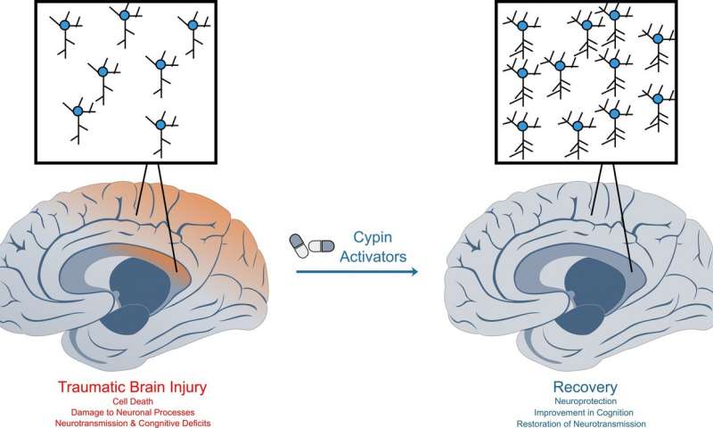
Traumatic brain injury causes widespread damage to neurons, leading to deficits in learning and memory. Cypin activators restore neuronal survival and function in mice, allowing for normal learning and memory. Credit: Mihir Patel/Rutgers University-New Brunswick

Traumatic brain injury: Discovery of two molecules could lead to new drug treatments

By Todd B. Bates, July 27, 2018, Rutgers University

After 10 years of research, a Rutgers-led team of scientists has identified two molecules that protect nerve cells after a traumatic brain injury and could lead to new drug treatments.

The molecules promote full recovery after traumatic brain injury (TBI) in mice, according to the study published online in Neurobiology of Disease. Traumatic brain injury is the leading cause of death for people under 45 years old in the United States and is associated with disability, early-onset dementia, cognitive disorders, mental illness and epilepsy.

Nearly all approaches for treating TBI focus on trying to prevent neurons, or nerve cells, from degenerating or on attempting to promote their survival, the study notes. TBI typically alters neural circuits within injured brain regions.

“The big issue with treatment after TBI is that there are no drugs that work well on patients to restore memory, and we’re targeting reconnectivity of neural circuitry,” said Bonnie L. Firestein, senior author of the study and a professor in the Department of Cell Biology and Neuroscience at Rutgers University-New Brunswick. “That means we want our neurons to function properly and connect with other neurons. We want to allow people to retain their cognition and ability to remember and learn, so our angle is novel.”

The researchers studied the protein cypin, an enzyme that breaks down guanine, which is an important building block for DNA and RNA in cells. The scientists previously showed that cypin is involved in promoting the proper shape in neurons and “keeping them happy,” Firestein said. This study found that speeding the breakdown of guanine protects neurons from injury and retains brain functioning.

Scientists at Rutgers-New Brunswick, University of Pennsylvania, Fox Chase Chemical Diversity Center Inc. and Columbia University want to develop drugs from the molecules for further studies.

Read the original article
 

With better devices, science can get closer to a more complete picture of how neurons interact for cognitive functionality. (Photo/iStock)

Are we getting closer to a complete brain mapping? New devices explore more regions safely

By Breanne Grady, April 13, 2018, viterbischool.usc.edu

Researchers have developed thin, flexible polymer-based materials that record activity in more subregions of the brain with safer, more specific placement.

Science has yet to unravel a complete understanding of the brain and all its intricate workings. It’s not for lack of effort.

Over many decades, multiple research studies have sought to understand the dizzying “talk,” or interconnectivity, between thousands of microscopic entities in the brain, in particular neurons. The goal: to one day arrive at a complete brain “mapping” — a feat that could unlock tremendous therapeutic potential.

Researchers at the USC Viterbi School of Engineering have developed thin, flexible polymer-based materials for use in microelectrode arrays that record activity more deeply in the brain and with more specific placement than ever before. What’s more is that each microelectrode array is made up of eight “tines,” each with eight microelectrodes which can record from a total 64 subregions of the brain at once.

Same Quality, More Safety

In addition, the polymer-based material, called Parylene C, is less invasive and damaging to surrounding cells and tissue than previous devices comprised of silicon or microwires. However, the long and thin probes can easily buckle upon insertion, making it necessary to add a dissolvable brace made up of polyethylene glycol (PEG) that prevents it from bending.

Professor Ellis Meng of the USC Viterbi Department of Biomedical Engineering said that the performance of the new polymer-based material is on par with microwires in terms of recording fidelity and sensitivity. “The information that we can get out is equivalent, but the damage is much less,” Meng said. “Polymers are gentler on the brain, and because of that, these devices get recordings of neuronal communication over long periods of time.”

As with any prosthetic implant, caution must be exercised in terms of the body’s natural immune response to a foreign element. In addition to inflammation, previous microelectrode brain implants made of silicon or microwires have caused neuronal death and glial scarring, which is damage to connective tissue in the nervous system. However, Parylene C is biocompatible and can be microfabricated in extremely thin form to mold well to specific subregions of the brain, allowing for exploration with minimal damage.

Listening In

So far, these arrays have been used to record synaptic responses of individual neurons within the hippocampus, a part of the brain responsible for memory formation. If injured, the hippocampus may be compromised, resulting in a patient’s inability to form new memories. Meng, a faculty member of the Michelson Center for Convergent Bioscience, said that the polymer-based material can conform to a specific location in the hippocampus and “listen in on a conversation” between neurons. Because there are many such “eavesdroppers” (the microelectrodes), much more information about their interconnectivity can be gleaned.

“I can pick where I want my electrodes to be, so I can match up to the anatomy of the brain,” Meng, the Dwight C. and Hildagarde E. Baum Chair, said. “Along the length of a tine, I can put a group of electrodes here and a group of electrodes there, so if we plant to a certain depth, it’s going to be near the neurons I want to record from.”

Up Next

Future research will determine the recording lifetime of polymer-based arrays and their long-term “signal-to-noise” (SNR) stability. Also, the team plans to create devices with even higher density, including a double-sided microelectrode array with 64 electrodes per tine instead of eight — making for a total of around 4,000 electrodes placed in the brain at once.

In addition to Meng, Professor Ted Berger, the David Packard Chair in Engineering, and Research Professor Dong Song (both of the USC Viterbi School of Engineering) were co-authors along with Ph.D. students Huijing Xu and Ahuva Weltman Hirschberg and post-doctoral scholar Kee Scholten. Funding was provided be the National Science Foundation (NSF) and the National Institutes of Health (NIH). The study titled “Acute in vivo testing of a conformal polymer microelectrode array for multi-region hippocampal recordings” now published in the Journal of Neural Engineering.

About the Michelson Center

The USC Michelson Center for Convergent Bioscience brings together a diverse network of premier scientists and engineers under one roof, thanks to a generous $50 million gift from orthopedic spinal surgeon, inventor and philanthropist Gary K. Michelson, and his wife, Alya Michelson. At the Michelson Center, scientists and engineers from the USC Dornsife College of Letters, Arts and Sciences, USC Viterbi School of Engineering and Keck School of Medicine of USC are working to solve some of the greatest intractable problems of the 21st century in biomedical science, including a fundamentally new understanding of the cell and new approaches for cancer, neurological and cardiovascular disease.

Read the original article
 

Serving the Brain Injury Community Since 1983