News

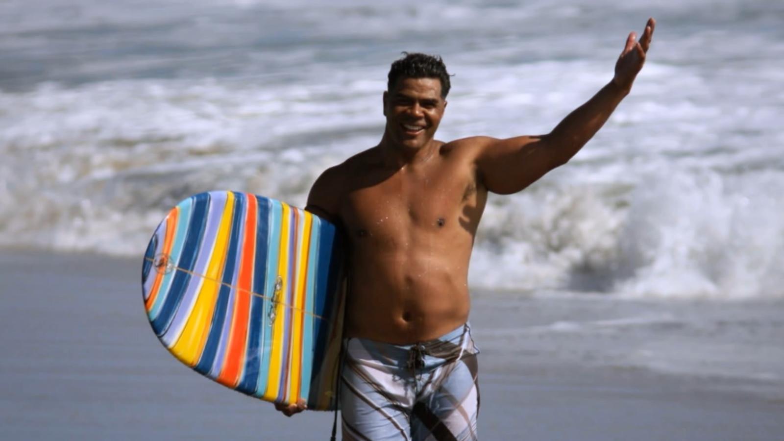
Junior Seau, shown at his beloved Pacific Ocean in the ESPN Films “30 for 30” documentary “Seau,” which premieres Thursday. (ESPN Films)

ESPN hits the mark with documentary ‘Seau’

By Tom Krasovic, September 20, 2018, San Diego Union Tribune

An aerial view of the Oceanside coast, in full sparkle and splendor below, grandly eases viewers into “Seau,” an ESPN Films documentary in the “30 for 30” series that debuts Thursday on the streaming service ESPN+.

It’s a sunny scene, the Pacific Ocean’s turquoise waves illuminated as they roll toward the white beach. The late Junior Seau told friends he found peace paddling on these waters, deep into his life alongside the town where he’d grown up.

Up at dawn with a yellow long-board and oar in hand, Seau had only a short walk from his beachfront home to the water.

Yet the former Chargers linebacker, role model and local philanthropist was then also writing in a journal of bouts with depression, memory loss and perceived guilt. There were headaches, too, and nights plagued by insomnia. “Buddy,” he’d told a friend and professional soccer player who’d suffered a brain injury from heading a ball, “I’ve had a concussion since I was 15.”

Diary entries also revealed feelings of humiliation and embarrassment over not living up to expectations of others and himself, and of feeling used by others.

“The world has nothing for me,” Seau pens in one entry, the cursive words all too legible.

One of Seau’s surviving adult children, after reading the grim line aloud, wonders why his father didn’t regard his family as something in this apparent world of nothing.

Why couldn’t they have been a lifeline for him to reach out and grasp?

“Seau,” produced and directed by Kirby Bradley, lets viewers draw their own conclusions about a complicated life that ended one May morning six years ago, at age 43, with a self-inflicted gunshot wound to the chest, but not before we hear from an array of family members, friends and experts in football and brain science.

At the end of the 90-minute film, themes of redemption and hope are raised.

“Let’s all walk from here being better for having known Junior Seau and the impact he had on our lives,” NFL quarterback Drew Brees, a former Chargers teammate of the Hall of Fame linebacker, concludes near the film’s end.

Former Chargers lineman Aaron Taylor notes that in death, Seau drew extraordinary attention to the link between head trauma and a degenerative brain disease, CTE, revealed in a tissue sample sent to a brain scientists at the family’s request.

Exciting beginnings and success are a thread to the film, followed often by bitter detours or hurtful endings.

Seau took to sports at Oceanside High with a passion that rivaled his stunning blend of size, speed and agility. If he was slamming into football ball-carriers or catching passes, scoring baskets or throwing the discus and shot, he was a “force of nature” for the green-and-white-clad Pirates, observers said.

A flood of football scholarship offers came to the small home where Seau and his brothers slept in a tiny garage.

Jubilation ensued when Seau chose USC, keeping him close to his parents and siblings and the tight-knit Samoan-American community in Oceanside. A similar celebration arose in 1990 when the Chargers drafted him fifth overall. “I’m a real momma’s boy,” Seau said, pulling on a blue team cap.

Playing for his beloved “Diego,” he led the long-struggling Chargers to the playoffs in just his third season, and their first Super Bowl two years later. “Now the world is gonna know the San Diego Chargers,” he told some 70,000 celebrants in Mission Valley after the team returned from claiming the 1994 AFC title in Pittsburgh.

The flip side?

If Oceanside lost a game in which he played, Junior lost his lunch money. It was the price his father exacted.

The thrill of signing with USC gave way to humiliation when a failed admittance test made him ineligible as a freshman. His father refused to talk to him in response, deeming the failure an embarrassment to the family. After a dominant junior year with USC, there would be no senior year. Making money was the next step, in no small part because he wanted to support his parents and other family members.

The Chargers couldn’t build upon their Super Bowl season, and the team’s constant losing wore on Seau.

When the Chargers traded him in the spring of 2003, after 13 seasons with the club, Seau was hurt that the team — Stay Unclassy, San Diego? — called not him but his agent to tell him the news. “I know that was hard on him,” said the agent, Steve Feldman.

Gina Seau was working for the Chargers in marketing when she first met Seau early in his NFL career.

She recalled “very kind eyes” and a “very soft voice” that almost “didn’t match the size and stature.”

The two would marry, but erratic behavior that Gina Seau linked to numerous football-related head injuries — “My head is on fire,” he told her — led to a divorce in 2002. The two remained friends. Believing that driving off a steep coastal cliff in October 2010 wasn’t an accident, Gina pleaded with her former husband to get help.

Here’s hoping that if there’s a “Seau II,” events yet to transpire bring more developments of redemption. Say, a cure for CTE.

Read the original article
 

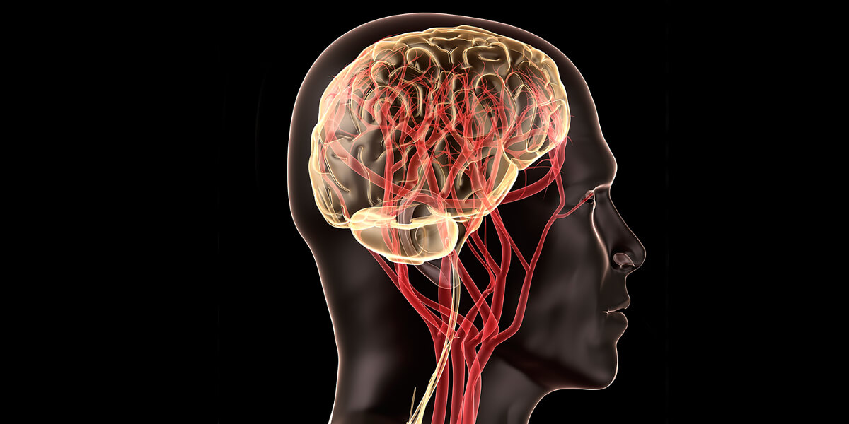
With better devices, science can get closer to a more complete picture of how neurons interact for cognitive functionality. (Photo/iStock)

Are we getting closer to a complete brain mapping? New devices explore more regions safely

By Breanne Grady, April 13, 2018, viterbischool.usc.edu

Researchers have developed thin, flexible polymer-based materials that record activity in more subregions of the brain with safer, more specific placement.

Science has yet to unravel a complete understanding of the brain and all its intricate workings. It’s not for lack of effort.

Over many decades, multiple research studies have sought to understand the dizzying “talk,” or interconnectivity, between thousands of microscopic entities in the brain, in particular neurons. The goal: to one day arrive at a complete brain “mapping” — a feat that could unlock tremendous therapeutic potential.

Researchers at the USC Viterbi School of Engineering have developed thin, flexible polymer-based materials for use in microelectrode arrays that record activity more deeply in the brain and with more specific placement than ever before. What’s more is that each microelectrode array is made up of eight “tines,” each with eight microelectrodes which can record from a total 64 subregions of the brain at once.

Same Quality, More Safety

In addition, the polymer-based material, called Parylene C, is less invasive and damaging to surrounding cells and tissue than previous devices comprised of silicon or microwires. However, the long and thin probes can easily buckle upon insertion, making it necessary to add a dissolvable brace made up of polyethylene glycol (PEG) that prevents it from bending.

Professor Ellis Meng of the USC Viterbi Department of Biomedical Engineering said that the performance of the new polymer-based material is on par with microwires in terms of recording fidelity and sensitivity. “The information that we can get out is equivalent, but the damage is much less,” Meng said. “Polymers are gentler on the brain, and because of that, these devices get recordings of neuronal communication over long periods of time.”

As with any prosthetic implant, caution must be exercised in terms of the body’s natural immune response to a foreign element. In addition to inflammation, previous microelectrode brain implants made of silicon or microwires have caused neuronal death and glial scarring, which is damage to connective tissue in the nervous system. However, Parylene C is biocompatible and can be microfabricated in extremely thin form to mold well to specific subregions of the brain, allowing for exploration with minimal damage.

Listening In

So far, these arrays have been used to record synaptic responses of individual neurons within the hippocampus, a part of the brain responsible for memory formation. If injured, the hippocampus may be compromised, resulting in a patient’s inability to form new memories. Meng, a faculty member of the Michelson Center for Convergent Bioscience, said that the polymer-based material can conform to a specific location in the hippocampus and “listen in on a conversation” between neurons. Because there are many such “eavesdroppers” (the microelectrodes), much more information about their interconnectivity can be gleaned.

“I can pick where I want my electrodes to be, so I can match up to the anatomy of the brain,” Meng, the Dwight C. and Hildagarde E. Baum Chair, said. “Along the length of a tine, I can put a group of electrodes here and a group of electrodes there, so if we plant to a certain depth, it’s going to be near the neurons I want to record from.”

Up Next

Future research will determine the recording lifetime of polymer-based arrays and their long-term “signal-to-noise” (SNR) stability. Also, the team plans to create devices with even higher density, including a double-sided microelectrode array with 64 electrodes per tine instead of eight — making for a total of around 4,000 electrodes placed in the brain at once.

In addition to Meng, Professor Ted Berger, the David Packard Chair in Engineering, and Research Professor Dong Song (both of the USC Viterbi School of Engineering) were co-authors along with Ph.D. students Huijing Xu and Ahuva Weltman Hirschberg and post-doctoral scholar Kee Scholten. Funding was provided be the National Science Foundation (NSF) and the National Institutes of Health (NIH). The study titled “Acute in vivo testing of a conformal polymer microelectrode array for multi-region hippocampal recordings” now published in the Journal of Neural Engineering.

About the Michelson Center

The USC Michelson Center for Convergent Bioscience brings together a diverse network of premier scientists and engineers under one roof, thanks to a generous $50 million gift from orthopedic spinal surgeon, inventor and philanthropist Gary K. Michelson, and his wife, Alya Michelson. At the Michelson Center, scientists and engineers from the USC Dornsife College of Letters, Arts and Sciences, USC Viterbi School of Engineering and Keck School of Medicine of USC are working to solve some of the greatest intractable problems of the 21st century in biomedical science, including a fundamentally new understanding of the cell and new approaches for cancer, neurological and cardiovascular disease.

Read the original article


The Cohen family partners with USC to serve families in Los Angeles.
 
by Lynn Lipinski, tfm.USC.edu (Autumn 2016) — PEACE AFTER WAR can be elusive for combat veterans who fight painful memories long after they’ve left the battlefield. Of the more than 2.6 million men and women who have served in the U.S. military since 9/11, about 20 percent experience some form of post-traumatic stress or brain injury—but nearly half forego treatment, according to the Cohen Veterans Network.

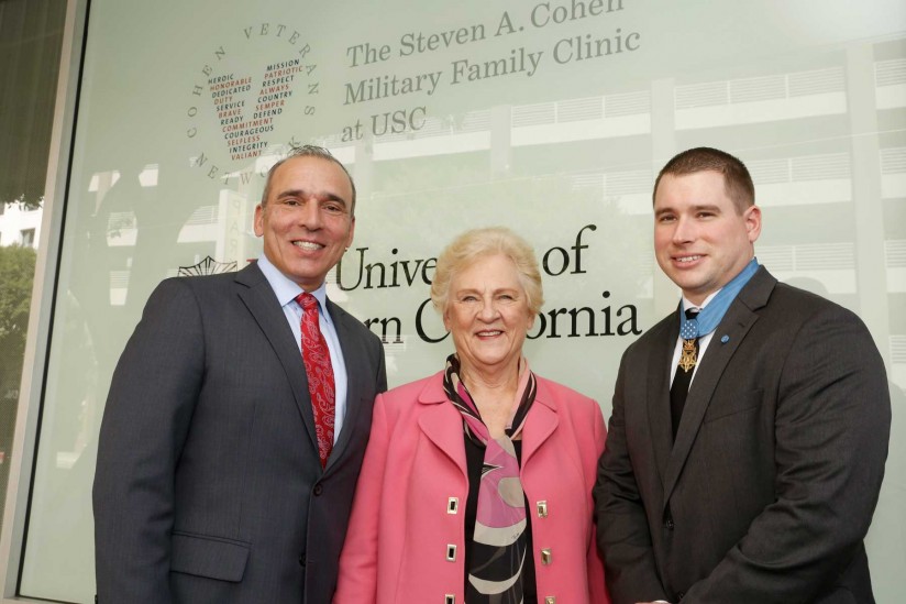
The Steven A. Cohen Military Family Clinic at USC, made possible by a $15.7 million gift from Steven Cohen and the Cohen Veterans Network, offers veterans and their family members free outpatient mental health services and case management. Recently opened in downtown Los Angeles, the Cohen Military Family Clinic at USC is part of a national network of clinics serving veterans and is a collaboration between the USC School of Social Work and the Keck School of Medicine of USC.

Providers will also be stationed at locations throughout the county in areas that otherwise lack these types of services. The clinic will also serve veterans who are ineligible for Veterans’ Admnistration benefits, such as those who served in the National Guard or the Reserves.

“The wounds of war are serious. It is not easy to serve your country in combat overseas and then come back into society seamlessly, especially if you are suffering,” says Cohen, chairman and CEO of Point72 Asset Management. “Veterans have paid an incredible price. It’s important that this country pays back that debt.”

The Cohen Veterans Network plans to create a system of about two dozen centers across the country by 2020 as part of a $275 million initiative to improve access to behavioral health care for recent veterans. Cohen’s support of services for veterans began in part because of a personal connection: His son, Robert, deployed to Afghanistan with the Marines and is currently in the Reserves.

USC’s strong programs for veterans made it a natural fit to host the clinic. The USC School of Social Work is home to the Center for Innovation and Research on Veterans and Military Families, where researchers conducted the first comprehensive study of veterans in L.A. County. Their findings are already helping to create effective services for veterans. The school has also earned national recognition for its pioneering master’s degree in military social work—the only program of its kind offered by a civilian research university.

Read the original article
 

When a person has a stroke, blood flow to the brain is interrupted, causing brain cells to die within minutes due to lack of oxygen. In some cases, this can result in paralysis, speech and language problems, vision problems, and memory loss. But in a new study, researchers have shown that stem cell therapy increases nerve cell production in mice with brain damage due to stroke.

by Marie Ellis, MedicalNewsToday.com (August 22, 2016) — The researchers – led by Berislav Zlokovic, M.D., Ph.D., from the University of Southern California (USC) – publish their findings in the journal Nature Medicine.

According to the Centers for Disease Control and Prevention (CDC), stroke is the fifth leading cause of death in the United States and is also a major cause of disability in adults.

The effects of a stroke depend on the location of the blockage and how much brain tissue is involved, but a stroke on one side of the brain will result in neurological effects on the opposite side of the body.

For example, a stroke on the right side of the brain could produce paralysis on the left side of the body, and vice versa.

A stroke in the brain stem can affect both sides of the body and could leave the patient in a so-called locked-in state, where the patient is unable to speak or move the body below the neck.

Given that about 800,000 people in the U.S. have a stroke each year, the researchers of this latest study wanted to investigate potential therapies.

Therapy is a combination of two methods

The researchers say their therapy is a combination of two methods. One involves surgically grafting human neural stem cells onto the damaged area, where they are able to mature into neurons and other brain cells.

The other therapy uses a compound called 3K3A-APC, which has been shown to help neural stem cells that have been grown in a petri dish grow into neurons. But the researchers say it was not clear what effect the molecule – called activated protein-C (APC) – would have on live animals.

As such, the team used mice for their experiment, and they found that a month after inducing stroke-like brain damage in the mice, those that had received both the stem cells and 3K3A-APC performed much better on motor and sensory function tests, compared with mice that received only one of the treatments or neither.

The researchers also observed that the mice given 3K3A-APC had more stem cells survive and mature into neurons.

But how did the researchers induce stroke-like brain damage in the mice? They disrupted blood flow to a specific brain area.

Then, 1 week later, which is the mouse equivalent of several months in humans, the researchers inserted the stem cells next to the dead tissue and administered either a placebo or 3K3A-APC.

“When you give these mice 3K3A-APC, it works much better than stem cells alone,” says Dr. Zlokovic. “We showed that 3K3A-APC helps the cells convert into neurons and make structural and functional connections with the host’s nervous system.”

‘No one in the stroke field has ever shown this’

The researchers also looked at the connections between the neurons that grew from the stem cells in the damaged brain region and nerve cells in the primary motor cortex.

The team found that the mice given the stem cells and 3K3A-APC had more neuronal connections – synapses – that linked those areas, compared with the mice given the placebo.

Then, when the researchers stimulated the mice’s paws with a vibration, the neurons that grew from the stem cells exhibited a stronger response in the mice that were treated.

“That means the transplanted cells are being functionally integrated into the host’s brain after treatment with 3K3A-APC. No one in the stroke field has ever shown this, so I believe this is going to be the gold standard for future studies.” ~Dr. Berislav Zlokovic

Following on from this study, the researchers want to pursue another phase II clinical trial to examine whether the treatment combination can encourage the growth of new neurons in human stroke patients to improve function.

They say that if that trial is successful, it could be possible to test the therapy’s effects on other conditions, including spinal cord injuries.

“This USC-led animal study could pave the way for a potential breakthrough in how we treat people who have experienced a stroke,” says Jim Koenig, Ph.D., program director at the National Institute of Health’s National Institute of Neurological Disorders and Stroke (NINDS), who funded the study.

“If the therapy works in humans,” he adds, “it could markedly accelerate the recovery of these patients.”

Read the original article

Serving the Brain Injury Community Since 1983