News

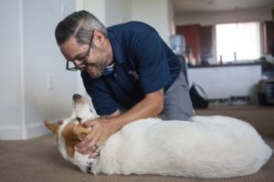
By John Prybys, LAS VEGAS REVIEW-JOURNAL (August 22, 2016) — Randy Dexter and Captain are more than just dog owner and dog. That’s obvious from the way Captain looks for Dexter whenever the Army veteran leaves the room, and the way the Lab mix’s demeanor slips instantly from playful to dead serious once he’s wearing the jacket that denotes his status as a service animal.

Dexter is a retired U.S. Army staff sergeant who did two tours of duty in Iraq. He was diagnosed with both post-traumatic stress disorder and a mild traumatic brain injury, and the story of Dexter and Captain is featured in a new awareness campaign urging veterans and military service people to seek help for traumatic brain injury if they need it.

The campaign, “A Head for the Future,” is sponsored by the Defense and Veterans Brain Injury Center. In his video, Dexter shares the struggles he has experienced coping with his injuries and the reluctance he felt at first to seek help for it.

But, he says, “I was lucky, because when I was in the Army and had my head injury, I was kind of forced to get help.”

Dexter, 34, is a graduate of Green Valley High School who served in the Army for 11 years and had two tours of duty in Iraq. In 2005, Dexter, a combat medic, and his squad were hit by an IED, prompting a long, and continuing, struggle with post-traumatic stress disorder.

Then, after returning home and while still being treated for PTSD and training soldiers bound for Iraq and Afghanistan, Dexter suffered a brain injury during a recreational football game. He’s not sure, even now, what happened. All he knows is that he lost the memory of about 24 hours’ time and, even, of going to the game at all.

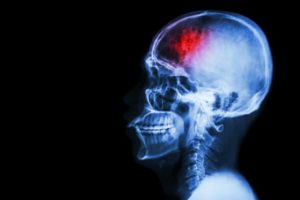
X-rays and imaging studies revealed no skull fractures or apparent injuries. But, afterward, Dexter experienced a worsening of already existing problems with his memory, concentration and equilibrium, and began to suffer migraines and severe, debilitating headaches that eventually compounded his PTSD and caused severe depression.

Dr. Scott Livingston, director of education for the Defense and Veterans Brain Injury Center in Silver Springs, Maryland, says symptoms of PTSD and brain injury often can overlap, making diagnosis a challenge. And when a brain injury does occur, he says, it often presents with no obvious symptoms that can be detected by X-ray or imaging scans.

In such cases, the problem likely is “more of a microscopic type of injury within the brain,” Livingston says.

Most civilians probably assume that brain injuries among service people are caused mostly by blasts and blunt-force trauma to the head. Yet, Livingston says, most are caused by motor vehicle collisions, training-related accidents, falls and sports and recreational activities.

Whatever the cause, military personnel are reluctant to report an injury or seek care for it. The current campaign is designed to raise awareness of brain injuries among service people, promote ways to prevent it when possible, and encourage men and women to report it and get it checked out, Livingston says.

“It’s well-known in scientific literature that the earlier someone reports a mild traumatic brain injury and goes to seek help, the better the chances are for better and more complete recovery,” he says.

During his treatment, Dexter participated in a program that paired injured veterans with service dogs. His experience with a dog named Ricochet was so good that he later welcomed the chance to be paired with Captain.

Dexter and Captain are a great team. Dexter says the dog can detect impending anxiety attacks even before he does, and that the dog can serve as a physical shield and protector in such public places as big-box retail stores, which can be particularly unnerving places for those with PTSD.

The true test of Captain’s effectiveness is that the dog has allowed Dexter to significantly reduce the medications he has to take. Today, it would be difficult for someone who doesn’t know the back story to detect Dexter’s struggles with traumatic brain injury, and it was his own previous interest in speaking out publicly about his conditions that led to his participation in the new awareness campaign.

Dexter now attends UNLV, where he’s majoring in communication studies and Spanish. He has been active on the debate teams, will be a peer adviser for other veterans, and hopes to kick off a music show on the university’s HD/internet radio station.

Dexter hopes his video and his story will help to persuade other veterans and active service people to seek out help for PTSD and brain injury. That can be difficult, he notes, because the standard soldier’s stance is that, whatever is happening, “you just deal with it, and that’s true across the whole military culture.”

Read the original article
 

By Alyssa Navarro, Tech Times (August 23, 2016) — Federal health regulators in the United States approved on Monday the use of two new computer softwares as cognitive screening tests for traumatic head injury patients.

Known as ImPACT or the Immediate Post-Concussion Assessment and Cognitive Testing (ImPACT), the new testing device, as well as a similar test designed for children, can be used by doctors to evaluate signs and symptoms of head injuries that could indicate concussion.

ImPACT is designed for patients aged 12 to 59 years old, while ImPACT Pediatric is intended for children aged 5 to 11 years old, officials said. Licensed health care professionals are the only ones allowed to perform the analysis and interpret the results.

The software can be accessed easily because it runs on both desktop computers and laptops, according to the U.S. Food and Drug Administration (FDA). Both tests the first ever devices permitted by the FDA to assess cognitive function after experiencing a possible concussion. They are designed to be part of medical evaluations in hospitals.

Although ImPACT and ImPACT Pediatric will definitely be useful for doctors, both tests are not meant to diagnose concussions or determine treatments that are appropriate for such cases, the FDA said.

Instead, both devices are only designed to test cognitive skills such as reaction time, memory and word recognition. All of these can be impacted by head injuries. Afterwards, the results are compared to a patient’s pre-injury baseline scores or an age-matched control database, the FDA said.

Dr. Carlos Peña, director of the neurological and physical medicine division at the Center for Devices and Radiological Health, acknowledges that the two testing devices can provide useful information that can aid doctors in the evaluation of people who are experiencing potential signs of concussion.

However, Peña says that clinicians should not completely depend on the tests alone to rule out concussion or to decide whether a player with a head injury should return to a game.

Statistics from the Centers for Disease Control and Prevention (CDC) reveal that traumatic brain injuries are responsible for more than 2 million visits to the emergency room in the country annually. Traumatic brain injuries also account for more than 50,000 deaths in America every year.

Cases of head injury among kids have been increasing. In May, a CDC report showed that from January 2001 to December 2013, approximately 214,883 children aged 14 years old and below were brought to emergency departments due to head injuries.

Read the original article
 

When a person has a stroke, blood flow to the brain is interrupted, causing brain cells to die within minutes due to lack of oxygen. In some cases, this can result in paralysis, speech and language problems, vision problems, and memory loss. But in a new study, researchers have shown that stem cell therapy increases nerve cell production in mice with brain damage due to stroke.

by Marie Ellis, MedicalNewsToday.com (August 22, 2016) — The researchers – led by Berislav Zlokovic, M.D., Ph.D., from the University of Southern California (USC) – publish their findings in the journal Nature Medicine.

According to the Centers for Disease Control and Prevention (CDC), stroke is the fifth leading cause of death in the United States and is also a major cause of disability in adults.

The effects of a stroke depend on the location of the blockage and how much brain tissue is involved, but a stroke on one side of the brain will result in neurological effects on the opposite side of the body.

For example, a stroke on the right side of the brain could produce paralysis on the left side of the body, and vice versa.

A stroke in the brain stem can affect both sides of the body and could leave the patient in a so-called locked-in state, where the patient is unable to speak or move the body below the neck.

Given that about 800,000 people in the U.S. have a stroke each year, the researchers of this latest study wanted to investigate potential therapies.

Therapy is a combination of two methods

The researchers say their therapy is a combination of two methods. One involves surgically grafting human neural stem cells onto the damaged area, where they are able to mature into neurons and other brain cells.

The other therapy uses a compound called 3K3A-APC, which has been shown to help neural stem cells that have been grown in a petri dish grow into neurons. But the researchers say it was not clear what effect the molecule – called activated protein-C (APC) – would have on live animals.

As such, the team used mice for their experiment, and they found that a month after inducing stroke-like brain damage in the mice, those that had received both the stem cells and 3K3A-APC performed much better on motor and sensory function tests, compared with mice that received only one of the treatments or neither.

The researchers also observed that the mice given 3K3A-APC had more stem cells survive and mature into neurons.

But how did the researchers induce stroke-like brain damage in the mice? They disrupted blood flow to a specific brain area.

Then, 1 week later, which is the mouse equivalent of several months in humans, the researchers inserted the stem cells next to the dead tissue and administered either a placebo or 3K3A-APC.

“When you give these mice 3K3A-APC, it works much better than stem cells alone,” says Dr. Zlokovic. “We showed that 3K3A-APC helps the cells convert into neurons and make structural and functional connections with the host’s nervous system.”

‘No one in the stroke field has ever shown this’

The researchers also looked at the connections between the neurons that grew from the stem cells in the damaged brain region and nerve cells in the primary motor cortex.

The team found that the mice given the stem cells and 3K3A-APC had more neuronal connections – synapses – that linked those areas, compared with the mice given the placebo.

Then, when the researchers stimulated the mice’s paws with a vibration, the neurons that grew from the stem cells exhibited a stronger response in the mice that were treated.

“That means the transplanted cells are being functionally integrated into the host’s brain after treatment with 3K3A-APC. No one in the stroke field has ever shown this, so I believe this is going to be the gold standard for future studies.” ~Dr. Berislav Zlokovic

Following on from this study, the researchers want to pursue another phase II clinical trial to examine whether the treatment combination can encourage the growth of new neurons in human stroke patients to improve function.

They say that if that trial is successful, it could be possible to test the therapy’s effects on other conditions, including spinal cord injuries.

“This USC-led animal study could pave the way for a potential breakthrough in how we treat people who have experienced a stroke,” says Jim Koenig, Ph.D., program director at the National Institute of Health’s National Institute of Neurological Disorders and Stroke (NINDS), who funded the study.

“If the therapy works in humans,” he adds, “it could markedly accelerate the recovery of these patients.”

Read the original article

  • 1
  • 2

Serving the Brain Injury Community Since 1983